. 2017 Dec 19;7(1):17864.
doi: 10.1038/s41598-017-17721-3.

Films of Bacteria at Interfaces (FBI): Remodeling of Fluid Interfaces by Pseudomonas aeruginosa

Affiliations

Films of Bacteria at Interfaces (FBI): Remodeling of Fluid Interfaces by Pseudomonas aeruginosa

Tagbo H R Niepa et al. Sci Rep. .

Erratum in

Abstract

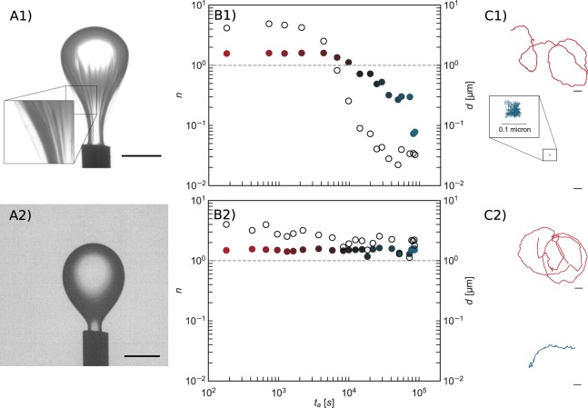
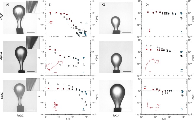
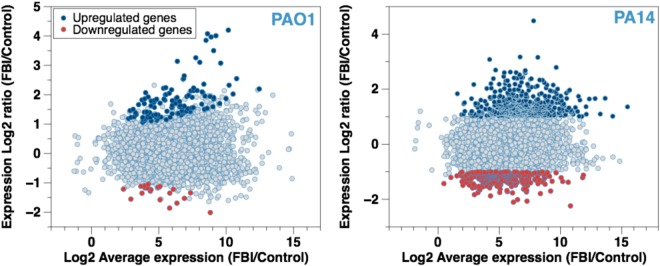
Bacteria at fluid interfaces endure physical and chemical stresses unique to these highly asymmetric environments. The responses of Pseudomonas aeruginosa PAO1 and PA14 to a hexadecane-water interface are compared. PAO1 cells form elastic films of bacteria, excreted polysaccharides and proteins, whereas PA14 cells move actively without forming an elastic film. Studies of PAO1 mutants show that, unlike solid-supported biofilms, elastic interfacial film formation occurs in the absence of flagella, pili, or certain polysaccharides. Highly induced genes identified in transcriptional profiling include those for putative enzymes and a carbohydrate metabolism enzyme, alkB2; this latter gene is not upregulated in PA14 cells. Notably, PAO1 mutants lacking the alkB2 gene fail to form an elastic layer. Rather, they form an active film like that formed by PA14. These findings demonstrate that genetic expression is altered by interfacial confinement, and suggest that the ability to metabolize alkanes may play a role in elastic film formation at oil-water interfaces.


Conflict of interest statement

The authors declare that they have no competing interests.

Figures

Figure 1
Figure 1
Films of bacteria at interfaces. (a) Low hexadecane toxicity for PAO1 and PA14 cells. Cells were re-suspended in saline solution (with or without a minimal media supplement, MMS) and exposed to hexadecane for 2 or 40 days and analyzed for viability by counting colony forming units (CFU) on an agar plate after incubation for 24 h. Cells remained viable for 40 days. (b) A PDMS platform with 50 micron diameter pores was fabricated to observe PAO1 and PA14 cells confined at the oil-water interface in the absence of nutrient. The cells display a differential response to the interface. PAO1 cells aggregate to form the “chef hat” structure. PA14 cells form an active layer characterized by a highly motile phase (scale bar: 20 μm). (c) FBI (Film of Bacteria at Interfaces) formation for P. aeruginosa PAO1 cells including the interaction of the bacteria with the oil droplet, adhesion, accumulation of the cells on the oil droplet, and maturation of the film. Single cells adhere to and cover the fluid interface over time. Interfaces are completely covered by a complex structure after 10 days (scale bar: 20 μm, 20 μm, and 1 mm, respectively) as cells adhere and accumulate as they secrete structures that form a film. (d) Scanning electron microscopy images of interfaces aged for 10 days reveal an asymmetric structure of PAO1 cells within a matrix of extracellular material (scale bar: 10 μm and 2 μm for low- and high-magnification images).
Figure 2
Figure 2
Characterization Of The Mechanics Of Bacteria Laden Interfaces. Panel (A): Pendant drop elastometry: a hexadecane droplet is held at the tip of an inverted needle in a bacteria suspension for 24 h and the drop surface is subsequently compressed by withdrawal of hexadecane. Scale bars: 1 mm. (A1) PAO1 laden interfaces are covered with a solid elastic film after 24 h. (A2) PA14 laden interfaces show no evidence of an elastic film formation over the same period. Panel (B): Exponent n (solid symbols) for lag times from 1.67 × 10−2–1.67 s and corresponding root mean square displacements d at lag time of 1.67 s (open circles) versus surface age. Color of the solid symbols, red to blue, indicates increased age. The dashed line, for reference, indicates the values of n and d of colloids subject to thermal Brownian motion in a bacteria-free hexadecane-water interface. (B1) PAO1 laden interfaces. (B2) PA14 laden interfaces. The PAO1 laden interface transitions to a nearly immobile film within 104 s. Probes at PA14 laden interface remain highly mobile for >104 s. Thereafter, d decays slightly as the interface becomes densely populated with bacteria, but the motion remains diffusive or super-diffusive. Panel (C): Typical trajectories traced by a colloidal probe for a 6 s time span at early (surface age of 60 seconds) and late (8.0 × 104 seconds) interface ages. Scale bars: 1 µm. (C1) PAO1 laden interfaces. (C2) PA14 laden interfaces. The colloid in the PAO1 laden interface is embedded in an elastic matrix.
Figure 3
Figure 3
Mutant Laden Interfaces. Panels (A & B): Pendant drop elastometry, exponent, n (solid circles), and root mean square displacement d (open circles), vs. interface age for PAO1ΔflgK, PAO1ΔpelA, and PAO1ΔpilC laden interfaces. The line at n = 100 indicates purely diffusive behavior; n < 10−1 indicates an essentially solid elastic film. The PAO1ΔflgK laden interface transforms from diffusive to elastic layers, while colloids at interfaces in contact with the other mutants (PAO1ΔpilC, and PAO1ΔpelA) remain superdiffusive and or diffusive indicating that they retain some fluid character over the course of the experiment. All PAO1 knockouts develop skins on pendant drops, evidenced by wrinkles upon compression. Insets to Panel (B): Typical trajectories traced by a colloidal probe over 3 s at bacteria laden interfaces at early (~102 seconds) and late (~8 × 104 seconds) interface ages. Scale bars, 1 micron, are the same for all trajectories. Panels (C & D): Pendant drop elastometry, exponent, n, and d, vs. interface age for PA14ΔflgK, PA14ΔpelA, and PA14ΔpilC laden interfaces; the PA14ΔpilC alone shows evidence of shear stress supporting film formation on the pendant drop; however, interfaces in contact with these mutants remain superdiffusive or diffusive through very late interface ages. Insets to panel (D): Typical trajectories traced by a colloidal probe over 3 s at bacteria laden interfaces at early (~102 seconds) and late (~8 × 104 seconds) interface ages. Scale bars, 1 micron, are the same for all trajectories.
Figure 4
Figure 4
MA (Mean-Average) Plot For Gene Expression Under FBI Conditions. Log2-fold of the change in expression vs. the Log2 average expression for each gene expressed by (a) PAO1 and (b) PA14 cells after confinement at a hexadecane-water interface for 1 h is presented. The total number of total mRNA expressed by the cells is plotted as a function of expression ratio. The blue and the red dots represent genes with significant changes in expression, by at least 2-fold with a false discovery rate (FDR) < 0.01.
Figure 5
Figure 5
Mechanics Of PAO1ΔalkB2 Laden Interfaces. Pendant drop compression (from left to right) performed on a droplet of hexadecane held in a suspension of PAO1ΔalkB2 for 24 h shows no evidence of elastic film formation; surface tension decreased until drop detached. Colloidal displacements remain superdiffusive for hours (lag times up to 1.67 s); the exponent n and RMS d only decay after 104 s; probe motion at late ages (~8 × 104 s) is significantly slowed and slightly subdiffusive, suggesting that the colloids are embedded in a weakly viscoelastic fluid interface rather than in a solid elastic film.

References

    1. Cohen TS, Prince A. Cystic fibrosis: a mucosal immunodeficiency syndrome. Nat. Med. 2012;18:509–519. doi: 10.1038/nm.2715. - DOI - PMC - PubMed
    1. Evans LR, Linker A. Production and characterization of the slime polysaccharide of Pseudomonas aeruginosa. J. Bacteriol. 1973;116:915–924. - PMC - PubMed
    1. Paradh, A. Gram-negative spoilage bacteria in brewing. Brewing Microbiology: Managing Microbes, Ensuring Quality and Valorising Waste, 175 (Woodhead Publishing, 2015).
    1. Wilking JN, Angelini TE, Seminara A, Brenner MP, Weitz DA. Biofilms as complex fluids. MRS bulletin. 2011;36:385–391. doi: 10.1557/mrs.2011.71. - DOI
    1. Wijman JG, de Leeuw PP, Moezelaar R, Zwietering MH, Abee T. Air-liquid interface biofilms of Bacillus cereus: formation, sporulation, and dispersion. Appl. Environ. Microbiol. 2007;73:1481–1488. doi: 10.1128/AEM.01781-06. - DOI - PMC - PubMed

Publication types

MeSH terms