. 2017 Dec 19;7(1):17788.
doi: 10.1038/s41598-017-17691-6.

Plasmon-actuated nano-assembled microshells

Affiliations

Plasmon-actuated nano-assembled microshells

Makiko T Quint et al. Sci Rep. .

Abstract

We present three-dimensional microshells formed by self-assembly of densely-packed 5 nm gold nanoparticles (AuNPs). Surface functionalization of the AuNPs with custom-designed mesogenic molecules drives the formation of a stable and rigid shell wall, and these unique structures allow encapsulation of cargo that can be contained, virtually leakage-free, over several months. Further, by leveraging the plasmonic response of AuNPs, we can rupture the microshells using optical excitation with ultralow power (<2 mW), controllably and rapidly releasing the encapsulated contents in less than 5 s. The optimal AuNP packing in the wall, moderated by the custom ligands and verified using small angle x-ray spectroscopy, allows us to calculate the heat released in this process, and to simulate the temperature increase originating from the photothermal heating, with great accuracy. Atypically, we find the local heating does not cause a rise of more than 50 °C, which addresses a major shortcoming in plasmon actuated cargo delivery systems. This combination of spectral selectivity, low power requirements, low heat production, and fast release times, along with the versatility in terms of identity of the enclosed cargo, makes these hierarchical microshells suitable for wide-ranging applications, including biological ones.


Conflict of interest statement

Methods and their use of nano-assembled microshells are the subject of patents and patent applications by the University of California Merced.

Figures
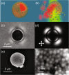
Figure 1
Figure 1
Characterization of a nano-assembled microshell (NAM). Schematic of (NAM) (a) An intact structure with sectional cut-out shows the encapsulated dye within. The wall is multiple layers thick. (b) Illumination by green light, resonant with the localized surface plasmon resonance (LSPR) of the nanoparticles in the wall, disrupts the structure due to photothermal heating, releasing the contents within. (c) Bright-field and (d) cross-polarized images of a NAM in liquid crystal medium. (d) SEM image of a NAM extracted from suspension and placed on an indium tin oxide coated glass slide. (e) Close-up of the same structure in (d), showing individual AuNPs that form the wall.
Figure 2
Figure 2
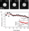
NAMs exhibit little to no leakage over many months. (ac) Fluorescence images of Lumogen F Red encapsulated in a NAM. (d) Dye intensity measured over five months. Inset: dye intensity encapsulated in shells compared to that of dye suspended in liquid crystal alone. The quantitative agreement between the two over the first ~10 days indicates that the small decrease is likely due to photo bleaching.
Figure 3
Figure 3
Actuation leading to release of contents from a NAM. (a) Fluorescence microscopy images of a NAM loaded with a fluorescent dye on a temperature-controlled stage. The temperature was increased from 80 to 108 °C, and the time after reaching 108 °C is given in the lower right corner. (b) Bright-field and (c) fluorescence time-lapse images during plasmon-actuated shell disintegration. The encapsulated dye is released during 5 s of illumination with 2 mW of incident power. Scale bars: 3 μm.
Figure 4
Figure 4
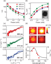
Spectral dependence of photothermal bubble formation. (a) The release time τ decreases with increasing power for three different excitation wavelengths; the fastest release is achieved at 514 nm, which is the wavelength closest to the LSPR (520 nm). (b) The equilibrium bubble radius R eq increases with increasing power, and is largest at 514 nm. (inset) cross-polarized image of the bubble shows isotropic phase inside and nematic phase outside. Scale bar: 3 μm. (c) The bubble radius r(t) increases over the first 100 ms of excitation at each wavelength. (d) Simulated thermal maps over a range of excitation wavelengths showing that photothermal temperature changes remain strongly localized to the NAM surface. Scale bar: 1 μm. (e) The extinction spectrum of a NAM with resonance at 520 nm (curve) shows good agreement with the maximum temperature change at the shell surface (filled circles).

References

    1. Yan Y, Warren SC, Fuller P, Grzybowski BA. Chemoelectronic circuits based on metal nanoparticles. Nat. Nanotechnol. 2016;11:603–608. doi: 10.1038/nnano.2016.39. - DOI - PubMed
    1. Polyak B, et al. Magnetic nanoparticle-mediated targeting of cell therapy reduces in-stent stenosis in injured arteries. ACS Nano. 2016;10:9559–9569. doi: 10.1021/acsnano.6b04912. - DOI - PubMed
    1. Tay A, Carlo DD. Magnetic nanoparticle-based mechanical stimulation for restoration of mechano-sensitive ion channel equilibrium in neural networks. Nano Lett. 2017;17:886–892. doi: 10.1021/acs.nanolett.6b04200. - DOI - PubMed
    1. Schein P, O’Dell D, Erickson D. Orthogonal nanoparticle size, polydispersity, and stability characterization with near-field optical trapping and light scattering. ACS Photonics. 2017;4:106–113. doi: 10.1021/acsphotonics.6b00628. - DOI
    1. Uchida T, Yoshikawa T, Tamura M, Iida T, Imura K. Multiple resonances induced by plasmonic coupling between gold nanoparticle trimers and hexagonal assembly of gold-coated polystyrene microspheres. J. Phys. Chem. Lett. 2016;7:3652–3658. doi: 10.1021/acs.jpclett.6b01493. - DOI - PubMed

Publication types