. 2017 Dec 19;7(1):17768.
doi: 10.1038/s41598-017-18125-z.

Analysis of killing of growing cells and dormant and germinated spores of Bacillus species by black silicon nanopillars

Affiliations

Analysis of killing of growing cells and dormant and germinated spores of Bacillus species by black silicon nanopillars

Sonali Ghosh et al. Sci Rep. .

Abstract

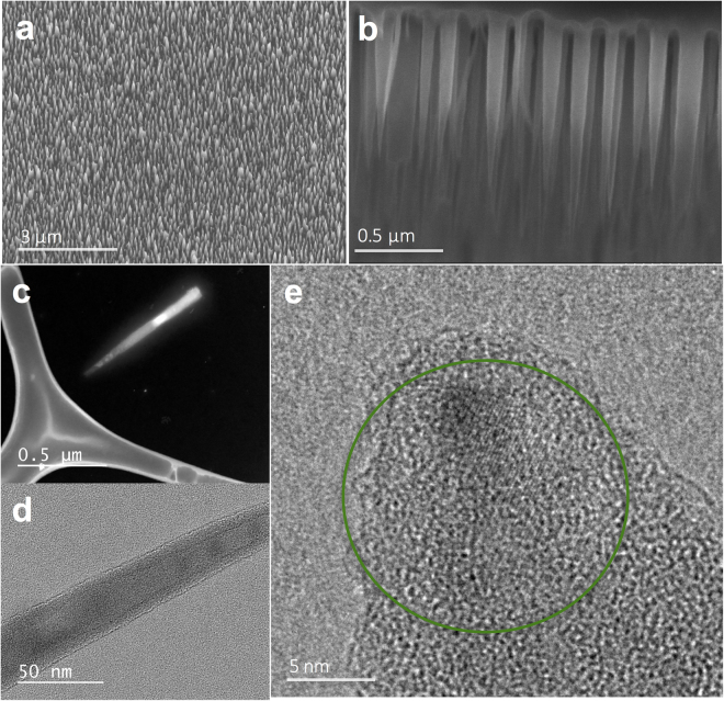
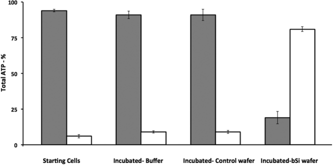
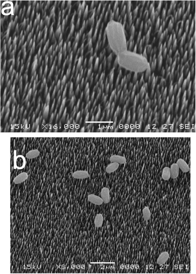
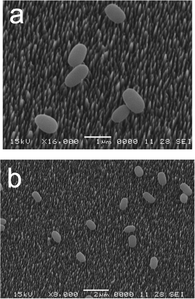
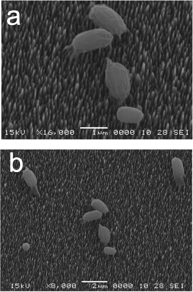
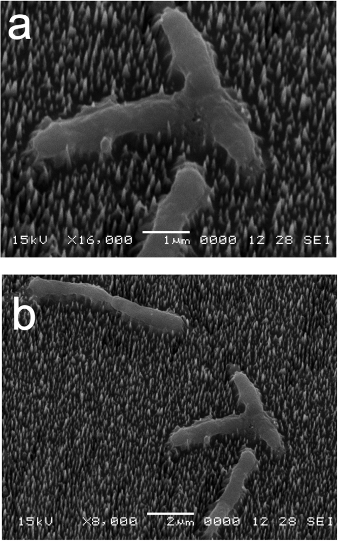
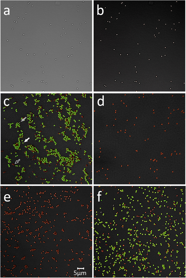
Black silicon (bSi) wafers with a high density of high-aspect ratio nanopillars have recently been suggested to have mechanical bactericidal activity. However, it remains unclear whether bSi with the nanopillars can kill only growing bacterial cells or also dormant spores that are harder to kill. We have reexamined the cidal activity of bSi on growing cells, dormant and germinated spores of B. subtilis, and dormant spores of several other Bacillus species by incubation on bSi wafers with and without nanopillars. We found that the bSi wafers with nanopillars were indeed very effective in rupturing and killing the growing bacterial cells, while wafers without nanopillars had no bactericidal effect. However, bSi wafers with or without nanopillars gave no killing or rupture of dormant spores of B. subtilis, Bacillus cereus or Bacillus megaterium, although germinated B. subtilis spores were rapidly killed. This work lays a foundation for novel bactericidal applications of bSi by elucidating the limits of mechanical bactericidal approaches.


Conflict of interest statement

The authors declare that they have no competing interests.

Figures

Figure 1
Figure 1
bSi wafers. (a) SEM image of as fabricated bSi wafer surface. (b) Cross-section SEM image of bSi nanopillars. (c) Dark field TEM imaging of one nanopillar on a specimen grid. (d,e) High resolution bright field TEM imaging of nanopillar body and tip structure with the green circle highlighting the crystalline regions, showing the core-shell nature of the bSi nanopillars.
Figure 2
Figure 2
ATP levels in pellet (gray bars) and supernatant (white bars) fractions of growing cells with or without incubation in buffer or on various black silicon wafers. Growing PS533 (wild-type) B. subtilis cells in PBS were prepared as described in Methods, and samples were incubated for 3 hr at 37 °C in buffer alone or on control or bSi wafers. Cells recovered from wafers, cells incubated at 37 °C and starting cells were centrifuged giving pellet (gray bars) and supernatant (white bars) fractions. These fractions were treated with boiling 1-propanol to extract ATP from intact cells and inactivate enzymes in supernatant fluid, and extracts were processed and assayed for ATP as described in Methods. All values shown are the percentages of total ATP in various samples, and are averages of assays of 3 samples; standard deviations for various values are also shown. Values for total ATP in 1 ml of various cell samples were between 8.4–9.4 pmol.
Figure 3
Figure 3
B. subtilis wild-type spores on a bSi wafer after a 48 hr incubation. (a,b) Spores of wild-type B. subtilis (PS533) were incubated on a bSi wafer with for 48 hr, the wafer dried, treated and electron micrographs were obtained as described in Methods.
Figure 4
Figure 4
B. subtilis coatless spores on a bSi wafer after a 48 hr incubation. (a,b) Spores of a severely coat-defective B. subtilis strain (PS4150) were incubated on a bSi wafer for 48 hr, the wafer dried, treated and electron micrographs were obtained as described in Methods.
Figure 5
Figure 5
B. cereus spores on a bSi wafer after a 48 hr incubation. (a,b) Spores of wild-type B. cereus were incubated on a bSi wafer for 48 hr, the wafer dried, treated and electron micrographs were obtained as described in Methods.
Figure 6
Figure 6
Growing B. subtilis wild-type cells on a bSi wafer with nanopillars after a 3 hr incubation. (a,b) Growing cells of wild-type B. subtilis (PS533) were incubated on a bSi wafer for 3 hr, the wafer dried, treated and electron micrographs were obtained as described in Methods.
Figure 7
Figure 7
Fluorescence micrographs of dormant and germinated B. subtilis spores stained with the BacLight reagent. B. subtilis PS533 (wild-type) spores, either dormant (panels a,b), or germinated (panels c–f) were incubated in various ways and then stained with the BacLight reagent and photographed by either differential contrast microscopy (panel a) or fluorescence microscopy (panels b–f) as described in Methods. Samples in panels c-f are: c) initial germinated spores; (d) initial germinated spores heated at 80 °C for 30 min; (e) germinated spores incubated on bSi wafers; and (f) spores incubated on control wafers. Arrows in panel c denote either a live spore staining green (closed arrow) a dead spore staining red (open arrow) or a dormant spore (dashed arrow). The scale bar in panel e is 5 μM, and the micrographs in all panels are at the same scale.

References

    1. Hasan J, Crawford RJ, Ivanova EP. Antibacterial surfaces: the quest for a new generation of biomaterials. Trends Biotechnol. 2013;31:295–304. doi: 10.1016/j.tibtech.2013.01.017. - DOI - PubMed
    1. Pogodin S, et al. Biophysical model of bacterial cell interactions with nanopatterned cicada wing surfaces. Biophys. J. 2012;104:835–840. doi: 10.1016/j.bpj.2012.12.046. - DOI - PMC - PubMed
    1. Hasan J, et al. Selective bactericidal activity of nanopatterned superhydrophobic cicada Psaltoda claripennis wing surfaces. Appl. Microbiol. Biotechnol. 2013;97:9257–9262. doi: 10.1007/s00253-012-4628-5. - DOI - PubMed
    1. Mainwaring DE, et al. The nature of inherent bactericidal activity: insights from the nanotopology of three species of dragonfly. Nanoscale. 2016;8:6527–6534. doi: 10.1039/C5NR08542J. - DOI - PubMed
    1. Ivanova EP, et al. Bactericidal activity of black silicon. Nature Commun. 2013;4:2838. doi: 10.1038/ncomms3838. - DOI - PMC - PubMed

Publication types