Review
. 2016 Oct 18:36:19.
doi: 10.1186/s41232-016-0025-2. eCollection 2016.

Liver regeneration and fibrosis after inflammation

Affiliations
Review

Liver regeneration and fibrosis after inflammation

Minoru Tanaka et al. Inflamm Regen. .

Abstract

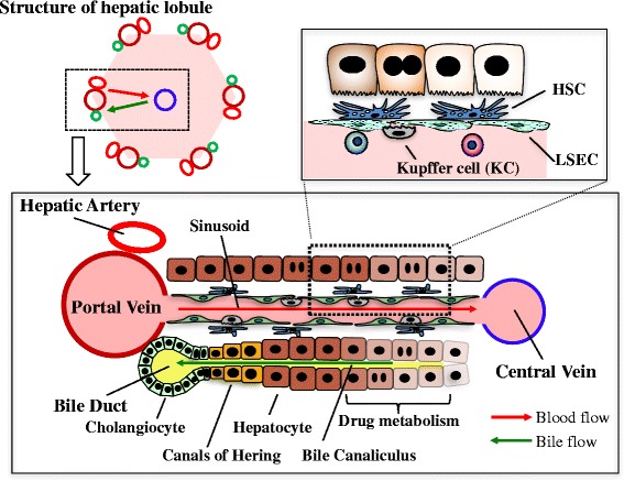
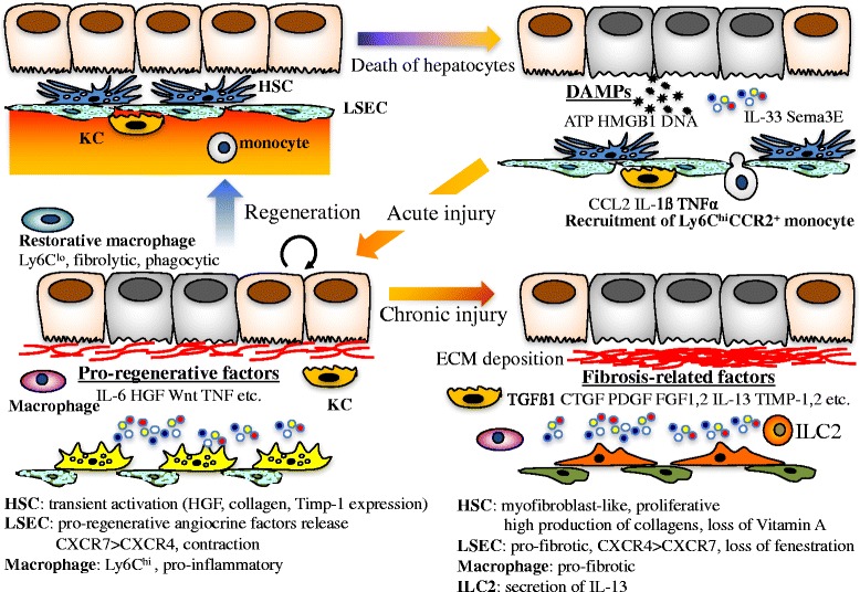
The liver is a unique organ with an extraordinary capacity to regenerate upon various injuries. In acute and transient liver injury by insults such as chemical hepatotoxins, the liver in rodents returns to the original architecture by proliferation and remodeling of the remaining cells within a week. In contrast, chronic liver inflammation due to various etiologies, e.g., virus infection and metabolic and immune disorders, results in liver fibrosis, often leading to cirrhosis and carcinogenesis. In both acute and chronic inflammation, a variety of immune and non-immune cells in the liver is involved in the processes resulting in either regeneration or fibrosis. In addition, chronic hepatitis often accompanies proliferation of atypical biliary cells, also known as liver progenitor cells or oval cells. Although the origin of liver progenitor cells and its contribution to hepatic repair is still under intense debate, recent studies have revealed a regulatory role for immune cells in progenitor proliferation and differentiation. In this review, we summarize recent studies on liver regeneration and fibrosis in the viewpoint of inflammation.

Keywords: Fibrosis; Hepatic stellate cell; Liver progenitor cell; Liver sinusoidal endothelial cell.


Figures

Fig. 1
Fig. 1
Schematic overview of the hepatic lobule. Blood flows into the liver from the portal vein and the hepatic artery toward the central vein through the sinusoid surrounded by fenestrated liver sinusoidal endothelial cells (LSECs). Bile produced by hepatocytes is collected into the bile ducts via the bile canaliculi surrounded by the apical membrane of hepatocytes. Kupffer cells (KC), resident macrophages in the liver, are located at the luminal side of the sinusoids, while hepatic stellate cells (HSCs) are positioned in close proximity to LSECs. The canals of Hering is the joint between hepatocytes and the bile ducts
Fig. 2
Fig. 2
Phenotypic changes of non-parenchymal cells associated with liver regeneration or fibrosis after injury

References

    1. Miyajima A, Tanaka M, Itoh T. Stem/progenitor cells in liver development, homeostasis, regeneration, and reprogramming. Cell Stem Cell. 2014;14:561–574. doi: 10.1016/j.stem.2014.04.010. - DOI - PubMed
    1. Suzuki A, Sekiya S, Onishi M, Oshima N, Kiyonari H, Nakauchi H, Taniguchi H. Flow cytometric isolation and clonal identification of self-renewing bipotent hepatic progenitor cells in adult mouse liver. Hepatology. 2008;48:1964–1978. doi: 10.1002/hep.22558. - DOI - PubMed
    1. Okabe M, Tsukahara Y, Tanaka M, Suzuki K, Saito S, Kamiya Y, Tsujimura T, et al. Potential hepatic stem cells reside in EpCAM+ cells of normal and injured mouse liver. Development. 2009;136:1951–1960. doi: 10.1242/dev.031369. - DOI - PubMed
    1. Higgins GM, Anderson RM. Experimental pathology of the liver, 1: restoration of the liver of the white rat following partial surgical removal. Arch Pathol. 1931;12:186–202.
    1. Miyaoka Y, Ebato K, Kato H, Arakawa S, Shimizu S, Miyajima A. Hypertrophy and unconventional cell division of hepatocytes underlie liver regeneration. Curr Biol. 2012;22:1166–1175. doi: 10.1016/j.cub.2012.05.016. - DOI - PubMed

LinkOut - more resources