. 2017 Nov 6:37:24.
doi: 10.1186/s41232-017-0054-5. eCollection 2017.

Effects of CTLA4-Ig on human monocytes

Affiliations

Effects of CTLA4-Ig on human monocytes

Toshihiro Tono et al. Inflamm Regen. .

Abstract

Background: Abatacept, a CTLA4-Ig fusion protein attenuates T cell activation by inhibiting the CD80/86-CD28 costimulatory pathway that is required for the proper T cell activation and thus displays beneficial effects in the treatment of rheumatoid arthritis (RA). Although some studies have disclosed the in vitro effects of this biological agent on the immune-competent cells, the precise mechanisms of action in RA still remain unclear. The current studies were therefore undertaken to explore the effects of abatacept on monocytes in detail.

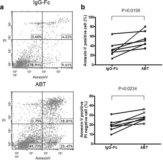
Methods: Monocytes from healthy donors were cultured in the presence of staphylococcal enterotoxin B (SEB) with pharmacologically attainable concentrations of abatacept or control IgG-Fc. The expression of CD80 and CD86 and the induction of apoptosis of monocytes were measured by flow cytometry. The expression of CD80 and CD86 messenger RNA (mRNA) was determined by quantitative RT-PCR.

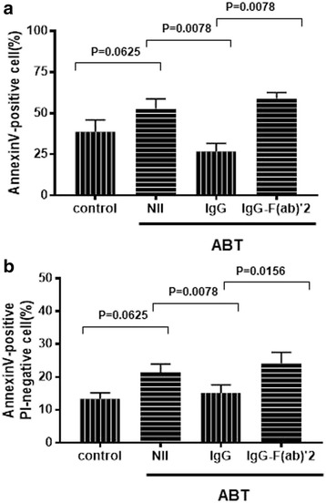
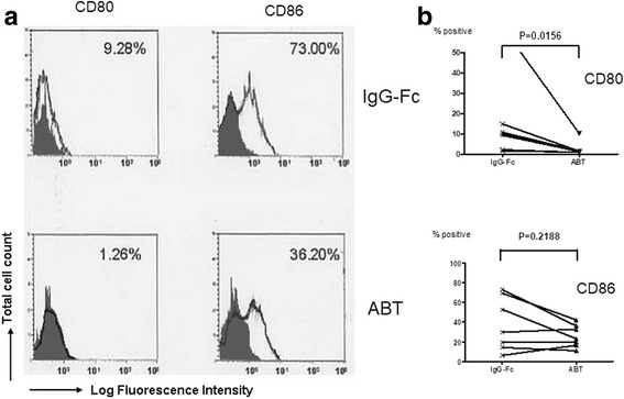
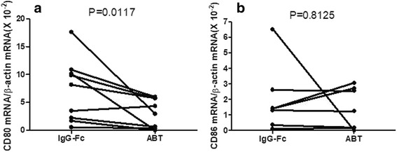
Results: Abatacept promoted apoptosis of SEB-stimulated monocytes. The induction of apoptosis of monocytes by these biological agents was reversed by the addition of IgG, but not IgG-F(ab')2 fragments. Furthermore, abatacept significantly suppressed the expression of CD80, but not that of CD86 at protein levels. Finally, abatacept significantly suppressed the expression of mRNA for CD80 of monocytes stimulated with SEB, but not that of CD86.

Conclusions: These results demonstrate that one of the mechanisms of action of abatacept involves the induction of apoptosis of monocytes, which involves interaction with Fc receptor on monocytes. Moreover, the data also demonstrate that abatacept selectively suppresses the expression of CD80 at mRNA levels.

Keywords: Abatacept; Apoptosis; Costimulation molecules; Monocytes.


Conflict of interest statement

Peripheral blood mononuclear cells (PBMCs) were obtained from healthy adult volunteers who gave informed consent. All procedures were approved by the ethics committee in the Kitasato University School of Medicine.Not applicable.The authors declare that they have no competing interests.Springer Nature remains neutral with regard to jurisdictional claims in published maps and institutional affiliations.

Figures

Fig. 1
Fig. 1
Effects of abatacept on annexin V expression of SEB-stimulated monocytes. Highly purified monocytes (1 × 106/well) from eight healthy individuals were cultured in the presence of SEB (100 pg/ml) in each well of 24-well flat-bottomed microtiter plates with abatacept (100 μg/ml) or control IgG-Fc (100 μg/ml) for 48 h, after which the cells were stained with FITC-conjugated annexin V and propidium iodide (PI). The cells were then analyzed by flow cytometry. a Representative dot plots for staining with FITC-conjugated annexin V and PI. b Percentages of total annexin V-positive cells (upper) or PI-negative annexin V-positive cells (lower) are shown. Statistical significance was evaluated by Wilcoxon’s signed-rank test
Fig. 2
Fig. 2
Differential effects of human IgG and IgG-F(ab′)2 fragments on the induction of apoptosis of SEB-stimulated monocytes by abatacept. Highly purified monocytes (1 × 106/well) from healthy individuals were cultured in the presence of SEB (100 pg/ml) in each well of 24-well flat-bottomed microtiter plates with abatacept (100 μg/ml) for 48 h, with or without IgG (100 μg/ml), or with IgG-F(ab′)2 fragments (100 μg/ml). After the cultures, the cells were stained with PE-conjugated annexin V, followed by analysis on flow cytometry. Percentages of total annexin V-positive cells (upper, n = 3) or PI-negative annexin V-positive cells (lower, n = 3) are shown. Error bars indicate SD values of three different experiments. Statistical significance was evaluated by Wilcoxon’s signed-rank test. V-positive cells (a) and PI-negative annexin V-positive cells (b)
Fig. 3
Fig. 3
Effects of abatacept on the expression of CD80 and CD86 on SEB-stimulated monocytes. Highly purified monocytes (1 × 106/well) from seven healthy individuals were cultured in the presence of SEB (100 pg/ml) in each well of 24-well flat-bottomed microtiter plates with abatacept (100 μg/ml) or control IgG-Fc (100 μg/ml) for 48 h, after which the cells were stained with FITC-conjugated anti-CD80, anti-CD86, or control IgG1, followed by counterstaining with PE-conjugated annexin V. The cells were then analyzed by flow cytometry. a Representative histograms of the staining of various molecules on annexin V-negative monocytes. The percentages of positive cells for specific mAb staining are indicated. Stainings with isotype-matched control mAb (control IgG1) are indicated by shade. b Percentages of positive cells for each specific mAb staining of monocytes from seven independent experiments are summarized. Statistical significance was evaluated by Wilcoxon’s signed-rank test
Fig. 4
Fig. 4
Effects of abatacept on the mRNA expression for CD80 and CD86 of SEB-stimulated monocytes. Highly purified monocytes (1 × 106/well) from healthy individuals were cultured in the presence of SEB (100 pg/ml) in each well of 24-well flat-bottomed microtiter plates with abatacept (100 μg/ml) or control IgG-Fc (100 μg/ml). After 24 h of incubation, the cells were harvested and the expression of mRNA for CD80 and CD86 were examined by quantitative RT-PCR. Data are expressed as the ratio to copy numbers of β-actin mRNA. Statistical significance was evaluated by Wilcoxon’s signed-rank test. CD80 (a) and CD86 (b)

References

    1. Korhonen R, Moilanen E. Abatacept, a novel CD80/86-CD28 T cell co-stimulation modulator, in the treatment of rheumatoid arthritis. Basic Clin Pharmacol Toxicol. 2009;104:276–284. doi: 10.1111/j.1742-7843.2009.00375.x. - DOI - PubMed
    1. Grohmann U, Bianchi R, Orabona C, Fallarino F, Vacca C, Micheletti A, et al. Functional plasticity of dendritic cell subsets as mediated by CD40 versus B7 action. J Immunol. 2003;171:2581–2587. doi: 10.4049/jimmunol.171.5.2581. - DOI - PubMed
    1. Munn DH, Sharma MD, Mellor AL. Ligation of B7-1/B7-2 by human CD4+ T cells triggers indoleamine 2,3-dioxygenase activity in dendritic cells. J Immunol. 2004;172:4100–4110. doi: 10.4049/jimmunol.172.7.4100. - DOI - PubMed
    1. Lahat N, Rahat MA, Ballan M, Cerem LW, Engelmayer M, Bitterman H. Hypoxia reduces CD80 expression on monocytes but enhances their LPS-stimulated TNF-α secretion. J Leukoc Biol. 2003;74:197–205. doi: 10.1189/jlb.0303105. - DOI - PubMed
    1. Mitoma H, Horiuchi T, Tsukamoto H, Tamimoto Y, Kimoto Y, Uchino A, et al. Mechanisms for cytotoxic effects of anti-tumor necrosis factor agents on transmembrane tumor necrosis factor alpha-expressing cells: comparison among infliximab, etanercept, and adalimumab. Arthritis Rheum. 2008;58:1248–1257. doi: 10.1002/art.23447. - DOI - PubMed

LinkOut - more resources