. 2017 Dec 8;5(1):e419.
doi: 10.1212/NXI.0000000000000419. eCollection 2018 Jan.

Phenotypic and functional complexity of brain-infiltrating T cells in Rasmussen encephalitis

Affiliations

Phenotypic and functional complexity of brain-infiltrating T cells in Rasmussen encephalitis

Faiez Al Nimer et al. Neurol Neuroimmunol Neuroinflamm. .

Abstract

Objective: To characterize the brain-infiltrating immune cell repertoire in Rasmussen encephalitis (RE) with special focus on the subsets, clonality, and their cytokine profile.

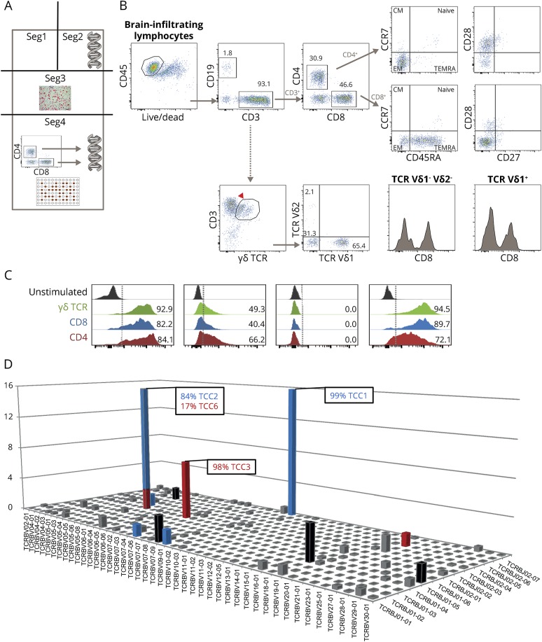
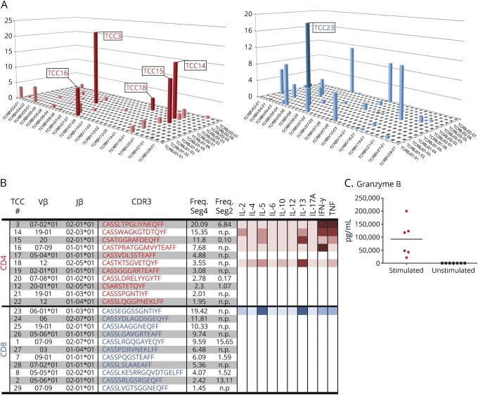
Methods: The immune cell infiltrate of freshly isolated brain tissue from RE was phenotypically and functionally characterized using immunohistology, flow cytometry, and T-cell receptor (TCR) deep sequencing. Identification of clonally expanded T-cell clones (TCCs) was achieved by combining flow cytometry sorting of CD4+ and CD8+ T cells and high-throughput TCR Vβ-chain sequencing. The most abundant brain-infiltrating TCCs were isolated and functionally characterized.

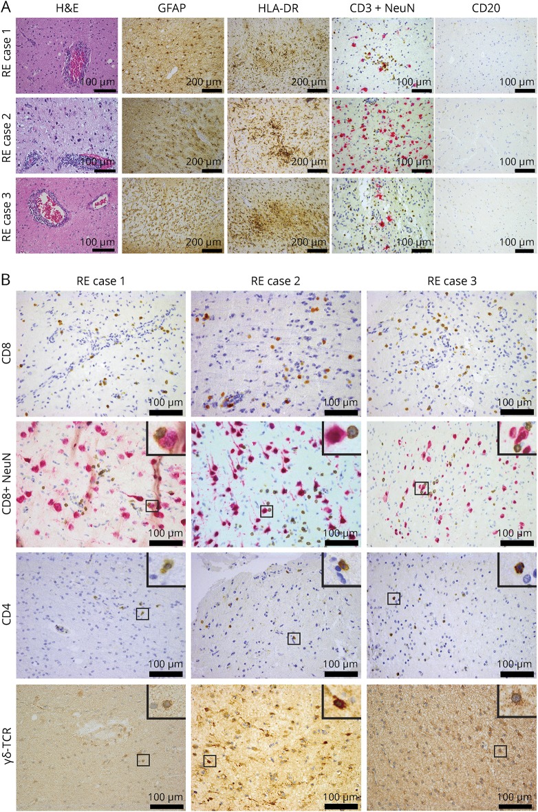
Results: We found that CD4+, CD8+, and also γδ T cells infiltrate the brain tissue in RE. Further analysis surprisingly revealed that not only brain-infiltrating CD8+ but also CD4+ T cells are clonally expanded in RE. All 3 subsets exhibited a Tc1/Th1 phenotype characterized by the production of interferon (IFN)-γ and TNF. Broad cytokine profiling at the clonal level showed strong production of IFN-γ and TNF and also secretion of interleukin (IL)-5, IL-13, and granzyme B, both in CD4+ and CD8+ T cells.

Conclusions: CD8+ T cells were until now considered the central players in the immunopathogenesis of RE. Our study adds to previous findings and highlights that CD4+ TCCs and γδ T cells that secrete IFN-γ and TNF are also involved. These findings underline the complexity of T-cell immunity in RE and suggest a specific role for CD4+ T cells in orchestrating the CD8+ T-cell effector immune response.


Figures

Figure 1
Figure 1. Coronal MRI images showing the evolution of white matter abnormality and atrophy of patient 1
MRI (fluid-attenuated inversion recovery, FLAIR) in February 2013 (A), in September 2013 (B), and after left vertical parasagittal hemispherotomy in October 2013 (C). The arrows in B show subcortical regions with white matter FLAIR signal abnormality. Note also the progressive atrophy of the brain and the left temporal lobe in particular.
Figure 2
Figure 2. Immunopathologic analysis of all 3 Rasmussen encephalitis cases
(A) Perivascular cuffing (hematoxylin and eosin [H&E]), prominent astrogliosis (GFAP), strong microglial activation (HLA-DR staining), T-cell infiltrates (CD3+NeuN), and absence of B cells (CD20) are shown. (B) Representative images showing CD8+ T cells infiltrating the tissue, some of them in proximity or direct contact to neurons (CD8+NeuN) and also, in all 3 cases, CD4+ T cells as well as γδ T cells (γδ-TCR) infiltrating the brain parenchyma. For CD8+NeuN, CD4, and γδ-TCR, a higher magnification of the small regions in squares is shown on the top right inset of each image. Scale bar = 100 μm except for GFAP and HLA-DR = 200 μm. GFAP = glial fibrillary acidic protein; HLA-DR = human leukocyte antigen; RE = Rasmussen encephalitis; TCR = T-cell receptor.
Figure 3
Figure 3. Methodological strategy, flow cytometric analysis, cytokine profile, and clonality of brain-infiltrating cells
(A) Methodological strategy of dissecting the brain tissue (dimensions: 2 × 6 × 1.5 cm) for various analyses: segment (seg) 1 was kept for further analyses. Seg2 was taken for DNA extraction and subsequent high-throughput TCR sequencing. Seg3 was embedded in paraffin and used in immunohistochemical studies. Seg4 was used for isolation of brain-infiltrating mononuclear cells with subsequent (1) phenotypic analyses by flow cytometry, (2) expansion with phytohemagglutinin, FACS of CD4+ and CD8+ T cells, DNA extraction, and subsequent high-throughput TCR sequencing, and (3) T-cell cloning (TCC). (B) Flow cytometry analysis (seg4) of the brain-infiltrating mononuclear cells. (C) Cytokine profile of brain-infiltrating T cells. (D) High-throughput TCR sequencing (seg2) showing oligoclonal expansions of both CD4+ and CD8+ T-cell infiltrates. CM = central memory; EM = effector memory; TCR = T-cell receptor.
Figure 4
Figure 4. Cytokine expression profile of individual T-cell clones
(A) The frequencies of CD4+ (red) or CD8+ (blue) TCCs that were identified in seg4 are shown in a 3D histogram. Note the similar clonality of CD4+ and CD8+ T cells. TCCs that were generated by limiting dilution and analyzed for cytokine expression are numbered and shown in bold. (B) The Vβ-chain, J-chain, and CDR3 sequence of TCRs from the most frequent TCCs as well as the cytokine profile of the derived TCCs are shown. Cytokine production is illustrated with a color gradient from pale to bolder that corresponds to lower and higher cytokine levels, respectively. (C) High levels of granzyme B secretion by both the CD4+ and CD8+ TCCs on stimulation. n.p. = not present. IFN = interferon; IL = interleukin; TCC = T-cell clone; TCR = T-cell receptor.

References

    1. Bien CG, Widman G, Urbach H, et al. . The natural history of Rasmussen's encephalitis. Brain 2002;125:1751–1759. - PubMed
    1. Rasmussen T, Olszewski J, Lloydsmith D. Focal seizures due to chronic localized encephalitis. Neurology 1958;8:435–445. - PubMed
    1. Mantegazza R, Bernasconi P, Baggi F, et al. . Antibodies against GluR3 peptides are not specific for Rasmussen's encephalitis but are also present in epilepsy patients with severe, early onset disease and intractable seizures. J Neuroimmunol 2002;131:179–185. - PubMed
    1. Bien CG, Schramm J. Treatment of Rasmussen encephalitis half a century after its initial description: promising prospects and a dilemma. Epilepsy Res 2009;86:101–112. - PubMed
    1. Jay V, Becker LE, Otsubo H, et al. . Chronic encephalitis and epilepsy (Rasmussen's encephalitis): detection of cytomegalovirus and herpes simplex virus 1 by the polymerase chain reaction and in situ hybridization. Neurology 1995;45:108–117. - PubMed