

A case of ophthalmomyiasis interna in the Pacific Northwest
- PMID: 29260045
- PMCID: PMC5722140
- DOI: 10.1016/j.ajoc.2017.01.002
A case of ophthalmomyiasis interna in the Pacific Northwest
Abstract
Purpose: We report a case of ophthalmomyiasis interna successfully removed in toto with pars plana vitrectomy.
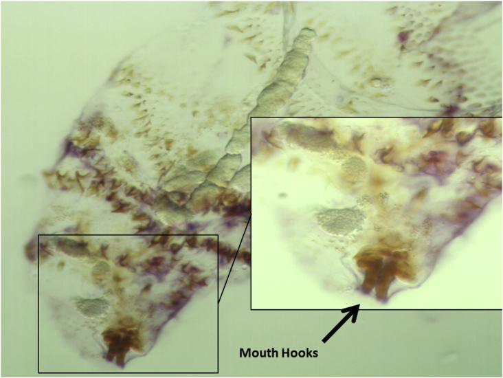
Observations: An 84-year-old woman with recent close contact with lambs presented with a new floater. Examination revealed subretinal tracks pathognomonic for ophthalmomyiasis and a larva suspended in the vitreous. The larva was successfully removed in toto with pars plana vitrectomy by aspiration through the vitreous cutter.
Conclusions and importance: Aspiration with pars plana vitrectomy can be considered a primary therapeutic modality for botfly larvae suspended in the vitreous. In our case, in toto removal of the larvae reduced the risk of inflammatory reaction.
Keywords: Oestrus ovis; Ophthalmomyiasis interna; Sheep botfly; Vitrectomy.
Figures

References
-
- Custis P.H., Pakalnis V.A., Klintworth G.K. Posterior interna ophthalmomyiasis; identification of a surgically removed Cuterebra larva by scanning electron microscopy. Ophthalmology. 1983;90:1583–1590. - PubMed
-
- O'Brien C.S., Allen J.H. Ophthalmomyiasis interna anterior: report of Hypoderma larva in anterior chamber. Am J Ophthalmol. 1939;22:996–998.
Publication types
LinkOut - more resources
Full Text Sources
Other Literature Sources
Miscellaneous
