

Optical coherence tomography angiography of iris microhemangiomatosis
- PMID: 29260048
- PMCID: PMC5722152
- DOI: 10.1016/j.ajoc.2017.02.003
Optical coherence tomography angiography of iris microhemangiomatosis
Abstract
Purpose: To report optical coherence tomography angiography (OCTA) of iris microhemangiomatosis.
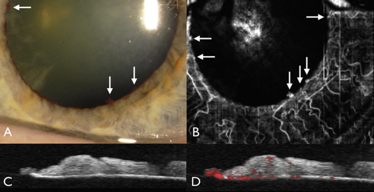
Observations: A 75-year-old asymptomatic Caucasian man was found to have bilateral pupillary vascular lesions during cataract evaluation. Visual acuity was counting fingers in the right eye (OD) and 20/40 in the left eye (OS) with normal intraocular pressures in both eyes (OU). In each eye there were multifocal, round, dark red, pinpoint vascular tufts at the pupillary margin, randomly distributed and numbering 1 in OD and 7 in OS, each measuring 0.2-0.3 mm in diameter and without active bleeding or hyphema. Fundus examination OU was normal. By fluorescein angiography, the multifocal pupillary vascular tufts demonstrated mild staining without leakage. By OCTA, the tufts were clearly delineated and were fed by normal appearing radial iris vessels. OCT b-scan documented the optically dense vascular tufts at 0.1 mm in thickness and angio-overlay confirmed blood flow emanating from the deep iris stroma. Observation was recommended with the option of cataract surgery to improve vision.
Conclusions and importance: Non-invasive imaging of iris microhemangiomatosis with OCTA delineates the vascular lesion with flow arising from the posterior iris stroma.
Keywords: Fluorescein angiography; Iris microhemangiomatosis; OCT; OCTA; Optical coherence tomography angiography.
Figures
References
-
- Shields C.L., Kancherla S., Patel J. Clinical survey of 3680 iris tumors based on patient age at presentation. Ophthalmology. 2012;119:407–414. - PubMed
-
- Shields J.A., Bianciotto C., Kligman B.E., Shields C.L. Vascular tumors of the iris in 45 patients: the 2009 Helen Keller lecture. Arch Ophthalmol. 2010;128:1107–1113. - PubMed
-
- Dahlmann A.H., Benson M.T. Spontaneous hyphema secondary to iris vascular tufts. Arch Ophthalmol. 2001;119:1728. - PubMed
-
- Goyal S., Foster P.J., Siriwardena D. Iris vascular tuft causing recurrent hyphema and raised IOP: a new indication for laser photocoagulation, angiographic follow-up, and review of laser outcomes. J Glaucoma. 2010;19:336–338. - PubMed
-
- Rosen E., Lyons D. Microhemangiomas at the pupillary border demonstrated by fluorescein photography. Am J Ophthalmol. 1969;67:846–885. - PubMed
Publication types
LinkOut - more resources
Full Text Sources
Other Literature Sources
