

The superficial and deep retinal capillary plexus in cases of fovea plana imaged by spectral-domain optical coherence tomography angiography
- PMID: 29260054
- PMCID: PMC5722189
- DOI: 10.1016/j.ajoc.2016.09.007
The superficial and deep retinal capillary plexus in cases of fovea plana imaged by spectral-domain optical coherence tomography angiography
Abstract
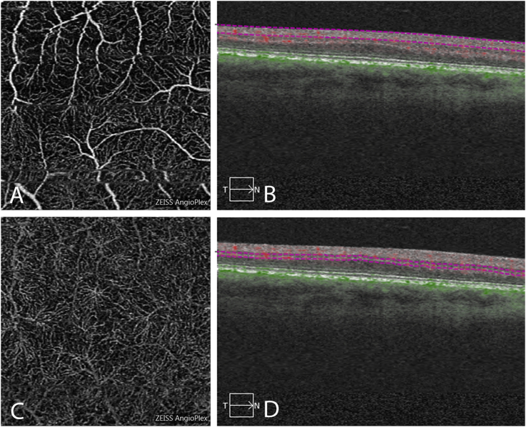
Purpose: To describe the appearance of the superficial and deep retinal capillary plexi in three patients with fovea plana of differing severity using spectral-domain optical coherence tomography angiography (OCTA).
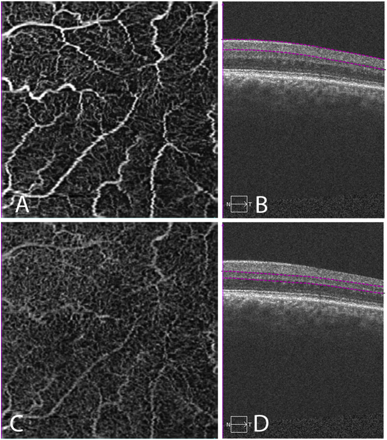
Observations: In the first case of grade 1 fovea plana (a patient with 20/25 vision), OCTA showed an orderly branching pattern of vessels from the superficial and deep retinal plexi extending to the center of the fovea. The second case of grade 3 fovea plana (20/30 vision) showed some disruption of the orderly vascular pattern with small caliber vessels from both superficial and deep layers densely covering the fovea center. Case 3 represented a patient with grade 4 fovea plana associated with PAX6 mutation and poor visual acuity. OCTA revealed a disorganized pattern of large and small caliber vessels from the superficial capillary network extending into the center of the fovea.
Conclusions and importance: Previously available imaging modalities were unable to specifically target different layers of the retinal vasculature. Using OCTA we have been able to show progressive changes in the vascular pattern in the deep and superficial retinal layers of patients with different grades of fovea plana. This novel imaging technique may play a role in the classification and assessment of patients with fovea plana.
Keywords: Fovea plana; Optical coherence tomography; Optical coherence tomography angiography; PAX6 mutation.
Figures

Similar articles
-
Volume Rendering of Angiographic Optical Coherence Tomography Angiography in Fovea Plana and Normal Foveal Pit.Front Neurol. 2021 Apr 27;12:633492. doi: 10.3389/fneur.2021.633492. eCollection 2021. Front Neurol. 2021. PMID: 33986716 Free PMC article.
-
Optical Coherence Tomography Angiography in Fovea Plana.Ophthalmic Surg Lasers Imaging Retina. 2016 Jul 1;47(7):670-3. doi: 10.3928/23258160-20160707-10. Ophthalmic Surg Lasers Imaging Retina. 2016. PMID: 27434900
-
Persistence of foveal capillary plexi in a case of fovea plana evident on OCT angiography.J Fr Ophtalmol. 2017 Jan;40(1):4-7. doi: 10.1016/j.jfo.2016.10.002. Epub 2016 Dec 15. J Fr Ophtalmol. 2017. PMID: 27989421
-
Assessment of macular microvasculature features before and after vitrectomy in the idiopathic macular epiretinal membrane using a grading system: An optical coherence tomography angiography study.Acta Ophthalmol. 2021 Nov;99(7):e1168-e1175. doi: 10.1111/aos.14753. Epub 2021 Jan 10. Acta Ophthalmol. 2021. PMID: 33423352
-
Quantitative evaluation of primary retinitis pigmentosa patients using colour Doppler flow imaging and optical coherence tomography angiography.Acta Ophthalmol. 2019 Nov;97(7):e993-e997. doi: 10.1111/aos.14047. Epub 2019 Apr 9. Acta Ophthalmol. 2019. PMID: 30963731
Cited by
-
Optical Coherence Tomography Angiography: Investigating Vessel Density Changes Induced by Caffeine in Healthy Subjects.J Ophthalmol. 2024 Oct 28;2024:5597188. doi: 10.1155/2024/5597188. eCollection 2024. J Ophthalmol. 2024. PMID: 39502492 Free PMC article.
-
Multimodal imaging in a patient with Prader-Willi syndrome.Int J Retina Vitreous. 2018 Nov 30;4:45. doi: 10.1186/s40942-018-0147-6. eCollection 2018. Int J Retina Vitreous. 2018. PMID: 30519487 Free PMC article.
-
Tilted disc in eyes with fovea plana.Graefes Arch Clin Exp Ophthalmol. 2023 Nov;261(11):3159-3164. doi: 10.1007/s00417-023-06161-7. Epub 2023 Jun 23. Graefes Arch Clin Exp Ophthalmol. 2023. PMID: 37351645
-
Evaluation of macular retinal oximetry across different levels of diabetic retinopathy: a cross sectional study.BMC Ophthalmol. 2025 Jan 17;25(1):24. doi: 10.1186/s12886-025-03850-1. BMC Ophthalmol. 2025. PMID: 39825268 Free PMC article.
-
Characterizing Foveal Hypoplasia Using Optical Coherence Tomography Angiography: Evaluation of Microvascular Abnormalities and Clinical Significance.J Clin Med. 2023 Jul 29;12(15):4992. doi: 10.3390/jcm12154992. J Clin Med. 2023. PMID: 37568394 Free PMC article.
References
-
- Meyer C.H., Lapolice D.J., Freedman S.F. Foveal hypoplasia in oculocutaneous albinism demonstrated by optical coherence tomography. Am J Ophthalmol. 2002;133(3):409–410. - PubMed
Publication types
LinkOut - more resources
Full Text Sources
Other Literature Sources
