Case Reports
. 2017 Jun 22:7:83-90.
doi: 10.1016/j.ajoc.2017.06.011. eCollection 2017 Sep.

Retinal findings in membranoproliferative glomerulonephritis

Affiliations
Case Reports

Retinal findings in membranoproliferative glomerulonephritis

Ahmad M Mansour et al. Am J Ophthalmol Case Rep. .

Abstract

Purpose: To assess the evolution of retinal findings in patients with membranoproliferative glomerulonephritis (MPGN) by funduscopy, intravenous fluorescein angiography and optical coherence tomography.

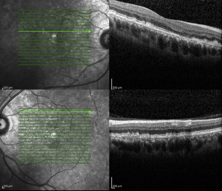
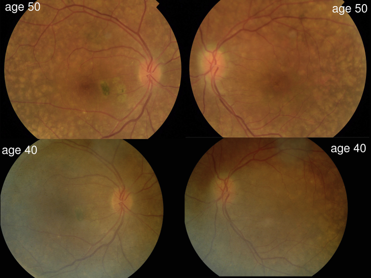
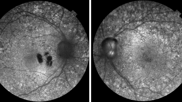
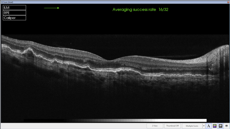
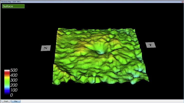
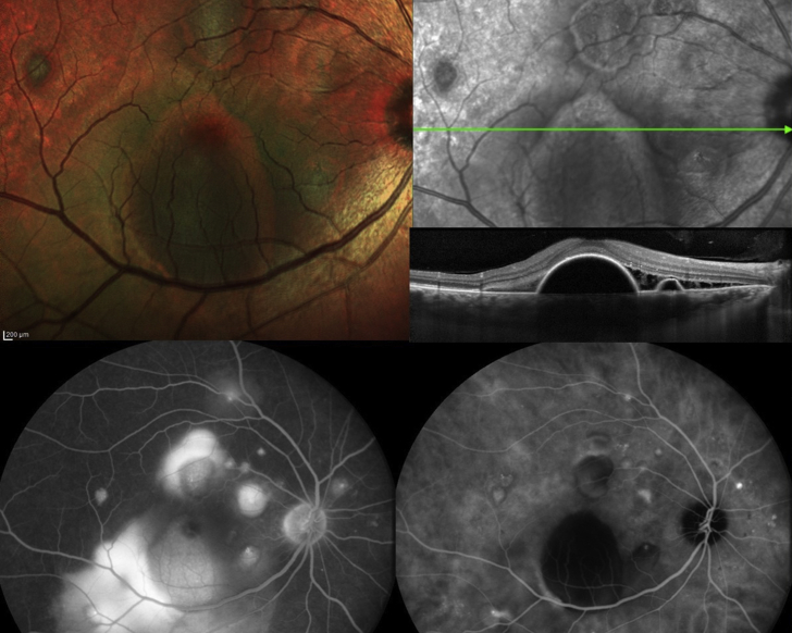
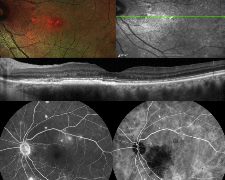
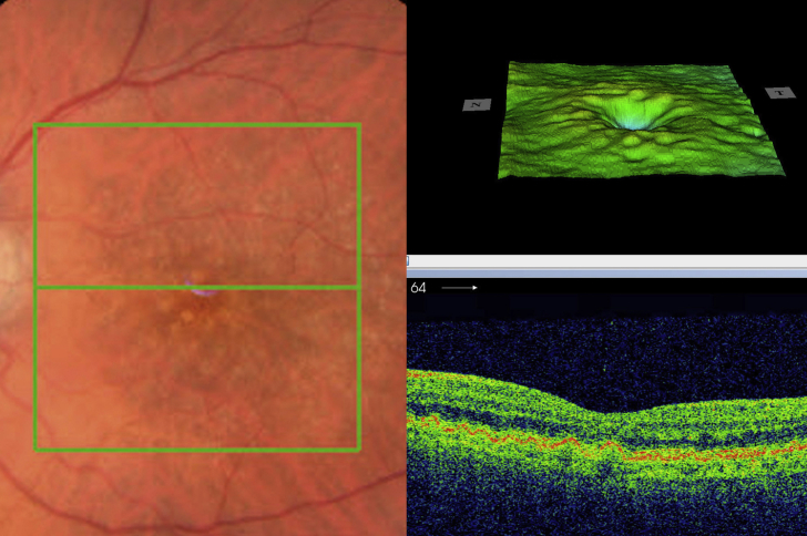
Observations: Three women and one man were followed for a period of 1.5-37 years. Four patients (8 eyes) had drusen detected at first fundus exam at age 24, 29, 50 and 55. Three patients (6 eyes) had diffuse thickening of Bruch's membrane, and two patients (3 eyes) had detachment of the retinal pigment epithelium with serous retinal detachment. Drusen tended to widen over a period of 10-year follow-up in one case.

Conclusions and importance: Drusen remain the ocular stigmata for MPGN occuring at an early age. The retinal disease is progressive with gradual thickening of Bruch's membrane and occurrence of retinal pigment epithelium detachment.

Keywords: Drusen; Macular degeneration; Membranoproliferative glomerulonephritis; Retinal pigment epithelial detachment.


Figures

Fig. 1
Fig. 1
Case 1-Color and autofluorescence of Case 1 showing posterior pole drusen in both eyes. (For interpretation of the references to colour in this figure legend, the reader is referred to the web version of this article.)
Fig. 2
Fig. 2
Case 1- Fluorescein angiography of the right eye and left eye shows hyperfluorescence of the drusen with drusen localized to the posterior pole (bottom left) and diseased retinal pigment epithelium (bottom right).
Fig. 3
Fig. 3
Case 1- Markedly thickened Bruch's membrane with elevation of retinal pigment epithelium at sites of drusen on Optical coherence tomography of both eyes.
Fig. 4
Fig. 4
Case 2- Color photographs of the posterior pole in both eyes showing enlargement of drusen over 10-year-follow-up. (For interpretation of the references to colour in this figure legend, the reader is referred to the web version of this article.)
Fig. 5
Fig. 5
Case 2- Autofluorescence showing bilateral diffuse stippled mottling of the retinal pigment epithelium, along with patches of sick retinal pigment epithelium in the right macula at the site of previous serous detachment of the retina.
Fig. 6
Fig. 6
Case 2- Diffuse thickening of Bruch's membrane with nodular elevations at sites of drusen on Optical coherence tomography of the right macula.
Fig. 7
Fig. 7
Case 2- Retinal pigment epithelium-internal limiting membrane Optical coherence tomography view of the left eye shows nodular elevations of retinal pigment epithelium corresponding to evenly spaced drusen.
Fig. 8
Fig. 8
Case 3- Fine drusen in the macular region in both eyes (Top) with no thickening of Bruch's membrane of the right macula (Bottom).
Fig. 9
Fig. 9
Case 4- Right posterior pole showing multiple detachment of retinal pigment epithelium (Top left), well outlined on Optical coherence tomography (Top right), with many pinpoint leakage noted on fluorescein angiography (Bottom left) and indocyanine green (Bottom right). (For interpretation of the references to colour in this figure legend, the reader is referred to the web version of this article.)
Fig. 10
Fig. 10
Case 4- Left posterior pole showing multiple alterations of retinal pigment epithelium with drusen (Top left) well outlined on Optical coherence tomography with diffuse thickening of Bruch's membrane (Top right); many pinpoint leakage sites are noted outside the arcade on fluorescein angiography (Bottom left) and indocyanine green (Bottom right). (For interpretation of the references to colour in this figure legend, the reader is referred to the web version of this article.)
Fig. 11
Fig. 11
(unpublished case)- This 79-year-old woman presented with visual loss from a right eye choroidal new vessel formation. The left macula had diffuse drusen (Left). Retinal pigment epithelium-internal limiting membrane view shows an evenly pitted pattern similar to case 2 (Right top). Optical coherence tomography (horizontal section) showed the density of the drusen by the dome-shaped even spaced elevations of the retinal pigment epithelium (Right bottom).

References

    1. Fakhouri F., Fremeaux-Bacchi V., Noel L.H. C3 glomerulopathy: a new classification. Nat Rev Nephrol. 2010;6:494–499. - PubMed
    1. Woo S.A., Ju H.Y., Kwon S.H. Reanalysis of membranoproliferative glomerulonephritis patients according to the new classification: a multicenter study. Kidney Res Clin Pract. 2014;33:187–191. - PMC - PubMed
    1. Dalvin L.A., Fervenza F.C., Sethi S., Pulido J.S. Manifestations of complement-mediated and immune complex-mediated membranoproliferative glomerulonephritis: a comparative consecutive series. Ophthalmology. 2016;123:1588–1594. - PubMed
    1. Dalvin L.A., Fervenza F.C., Sethi S., Pulido J.S. Shedding light on fundus drusen associated with membranoproliferative glomerulonephritis: breaking stereotypes of types I, II, and III. Retin Cases Brief Rep. 2016;10:72–78. - PubMed
    1. Polk T.D., Kimura A.E., Park D., Gass J.D. Subretinal fluid associated with membranoproliferative glomerulonephritis. Arch Ophthalmol. 1997;115:927–928. - PubMed

Publication types

LinkOut - more resources