

Recovery of outer retinal laminations on optical coherence tomography after treatment of cancer associated retinopathy
- PMID: 29260107
- PMCID: PMC5731551
- DOI: 10.1016/j.ajoc.2017.08.001
Recovery of outer retinal laminations on optical coherence tomography after treatment of cancer associated retinopathy
Abstract
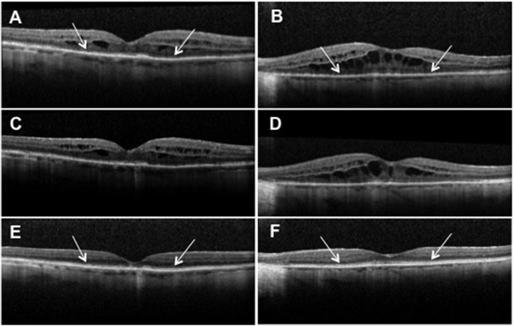
Purpose: To report novel optical coherence tomography findings in a case of anti-α-enolase cancer associated retinopathy.
Observations: An elderly female presented with bilateral decreased vision and a recent diagnosis of ovarian carcinoma. Optical coherence tomography demonstrated bilateral loss of outer retinal structures and macular edema. Serum testing found antibodies against α-enolase and 82-84 kDa proteins. Outer retinal structures showed recovery, macular edema resolved and repeat anti-retinal antibody testing became negative following cancer therapy and topical difluprednate treatment.
Conclusions and importance: Cancer associated retinopathy is a paraneoplastic disease that results in damage to retinal structures through an autoimmune response. The damage is generally considered to be irreversible; however, in rare cases, such as observed here, retinal structures may demonstrate recovery after treatment.
Keywords: Cancer associated retinopathy; Optical coherence tomography.
Figures
References
-
- Rahimy E., Sarraf D. Paraneoplastic and non-paraneoplastic retinopathy and optic neuropathy: evaluation and management. Surv Ophthalmol. 2013;58(5):430–458. - PubMed
-
- Weleber R.G., Watzke R.C., Shults W.T. Clinical and electrophysiologic characterization of paraneoplastic and autoimmune retinopathies associated with anti-enolase antibodies. Am J Ophthalmol. 2005;139(5):780–794. - PubMed
-
- Magrys A., Anekonda T., Ren G., Adamus G. The role of anti-alpha-enolase autoantibodies in pathogenicity of autoimmune-mediated retinopathy. J Clin Immunol. 2007;27(2):181–192. - PubMed
Publication types
Grants and funding
LinkOut - more resources
Full Text Sources
Other Literature Sources
