

doi: 10.3201/eid2401.170401.
Serologic Evidence of Fruit Bat Exposure to Filoviruses, Singapore, 2011-2016
- PMID: 29260678
- PMCID: PMC5749470
- DOI: 10.3201/eid2401.170401
Item in Clipboard
Serologic Evidence of Fruit Bat Exposure to Filoviruses, Singapore, 2011-2016
Emerg Infect Dis.
2018 Jan.
Abstract
To determine whether fruit bats in Singapore have been exposed to filoviruses, we screened 409 serum samples from bats of 3 species by using a multiplex assay that detects antibodies against filoviruses. Positive samples reacted with glycoproteins from Bundibugyo, Ebola, and Sudan viruses, indicating filovirus circulation among bats in Southeast Asia.
Keywords: Bundibugyo virus; Ebola virus; Singapore; Southeast Asia; Sudan virus; filoviruses; fruit bats; serology; virus envelope glycoprotein; virus surveillance; viruses.
Figures
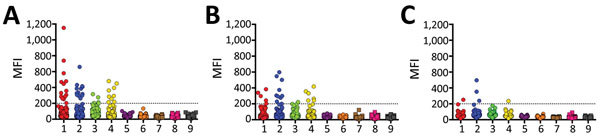
Mean fluorescence intensity (MFI) values obtained from Bio-Plex assay (Bio-Rad, Hercules, CA, USA) screening of individual serum samples from bats of 3 species with soluble filovirus glycoproteins. Dashed line indicates the cutoff value at 200 MFI. 1, Zaire ebolavirus; 2, Bundibugyo ebolavirus; 3, Taï Forest ebolavirus; 4, Sudan ebolavirus; 5, Reston ebolavirus–monkey; 6, Reston ebolavirus–pig; 7, Marburg virus–Musoke; 8, Marburg virus–Angola; 9, Ravn virus.
Western blot results of individual bat serum samples probed against Zaire ebolavirus and Bundibugyo ebolavirus glycoproteins 1 and 2 (GP1, GP2). Boldface indicates positivity by Western blot and underlining indicates positivity by Bio-Plex (Bio-Rad, Hercules, CA, USA). 1, soluble GP1 and GP2 blotted with control anti–Ebola virus nonhuman primate polyclonal serum that demonstrates cross-reactivity with Bundibugyo ebolavirus soluble GP. Other numbers along baseline correspond to the following sample identifiers, also used in Table 2: 2, 0805149; 3, 012309; 4, 011603; 5, 0116048; 6, 0719036; 7, 1128015; 8, 0726122; 9, 042701; 10, 040807; 11, 0512540; 12, 1009010; 13, 0408029; 14, 070409; 15, 112112; 16, 062590; 17, 0228004; 18, 0919025; 19, 0625095. BDBV, Bundibugyo virus; EBOV, Ebola virus.
References
Publication types
MeSH terms
Substances
LinkOut - more resources
Full Text Sources
Other Literature Sources
Medical
