Case Reports

Characterization of a Feline Influenza A(H7N2) Virus

Masato Hatta et al. Emerg Infect Dis. 2018 Jan.

Abstract

During December 2016-February 2017, influenza A viruses of the H7N2 subtype infected ≈500 cats in animal shelters in New York, NY, USA, indicating virus transmission among cats. A veterinarian who treated the animals also became infected with feline influenza A(H7N2) virus and experienced respiratory symptoms. To understand the pathogenicity and transmissibility of these feline H7N2 viruses in mammals, we characterized them in vitro and in vivo. Feline H7N2 subtype viruses replicated in the respiratory organs of mice, ferrets, and cats without causing severe lesions. Direct contact transmission of feline H7N2 subtype viruses was detected in ferrets and cats; in cats, exposed animals were also infected via respiratory droplet transmission. These results suggest that the feline H7N2 subtype viruses could spread among cats and also infect humans. Outbreaks of the feline H7N2 viruses could, therefore, pose a risk to public health.

Keywords: H7N2; New York; United States; feline; influenza; influenza virus; respiratory infections; viruses; zoonoses; zoonotic infection.


Figures

Figure 1
Figure 1
Phylogenetic tree of influenza A viral HA segments. Phylogenetic analysis was performed for selected influenza A viruses representing major lineages. The evolutionary history was inferred using the neighbor-joining method (12). The optimal tree with the branch length sum of 1.22521320 is shown. The percentage of replicate trees in which the associated taxa clustered together in the bootstrap test (500 replicates) is shown next to the branches (13). The tree is drawn to scale, with branch lengths in the same units as those of the evolutionary distances used to infer the phylogenetic tree. The evolutionary distances were computed using the Tamura 3-parameter method (14) and are in the units of the number of base substitutions per site. The analysis involved 44 nt sequences. Codon positions included were 1st + 2nd + 3rd + noncoding. All positions containing gaps and missing data were eliminated. The final dataset contained a total of 1,612 positions. Evolutionary analyses were conducted in MEGA7 (15).
Figure 2
Figure 2
Growth properties of A/feline/NY/16 and A/chicken/NY/99 viruses in mammalian and avian cells at different temperatures. Cells were infected with viruses at a multiplicity of infection of 0.005 and incubated at 33° and 37°C (or at 37°C and 39°C for avian CEF cells). Supernatants were harvested at the indicated time points. Virus titers were determined by use of plaque assays in Madin-Darby canine kidney (MDCK) cells. A549, human lung carcinoma epithelial cells; Clone81, cat kidney fibroblast cells; Fc2Lu, cat lung cells; CEF, chicken embryo fibroblast cells. The species from which the cell lines are derived are shown. The values presented are the averages of 3 independent experiments ±standard deviation. Statistical significance was determined as described in the Technical Appendix. *, p<0.05; **, p<0.01; ***, p<0.001; ****, p<0.0001.
Figure 3
Figure 3
Body weight and temperature changes in cats infected with A/feline/NY/16 and A/chicken/NY/99 viruses. Three cats per group were infected intranasally with 106 PFU of viruses and monitored for bodyweight and temperature changes.
Figure 4
Figure 4
Immunohistochemistry findings in infected cats. Shown are representative sections of nasal turbinates and lungs of cats infected with the indicated viruses on days 3 and 6 postinfection. Three cats per group were infected intranasally with 106 PFU of virus, and tissues were collected on days 3 and 6 postinfection. Type A influenza virus nucleoprotein (NP) was detected by a mouse monoclonal antibody to this protein. For nasal turbinate sections, -: no NP-positive cells, +/−: NP-positive cells detected in 1–2 focal regions, +: NP-positive cells detected in more than two focal regions, 2+: NP-positive cells in large regions. For bronchus and alveolar sections, -: no NP-positive cells, +/−: Up to 5 NP-positive cells, +: Six or more NP-positive cells. NP-positive cells were detected in focal, but not in diffuse bronchial and alveolar sections. For all analyses, the entire sections were evaluated. Scale bars, 50 μm (nasal turbinates), 100 μm (lung).
Figure 5
Figure 5
Pathology findings in cats infected with A/feline/NY/16 influenza A(H7N2) virus on day 6 postinfection, New York, NY, USA. A) In lungs, moderately severe histopathologic changes are present in the lower airways. The lamina propria of bronchi (B) and bronchioles (Br) and the surrounding interstitium are infiltrated by numerous histiocytes, lymphocytes, and plasma cells (*), which also extend into and expand neighboring alveolar septa. The infiltrates extend into and expand nearby alveolar septa. The lumina of bronchioles are filled with numerous foamy macrophages, viable and degenerating neutrophils, proteinaceous fluid, and sloughed respiratory epithelial cells. Hyperplasia of bronchiole-associated lymphoid tissue (open arrow) and perivascular edema (solid arrow) are present. Scale bar indicates 500 µm. B) In nasal cavities, copious amounts of exudate are present comprising numerous degenerating and necrotic neutrophils, cellular debris, proteinaceous fluid, and strands of mucin. The respiratory epithelium covering the nasal turbinates (T) is extensively eroded. The underlying lamina propria appears diffusely bluish-purple due to infiltration by moderate-to-large numbers of histiocytes, neutrophils, lymphocytes, and plasma cells (*). Scale bar indicates 500 µm. C) In the trachea, a locally extensive focus of inflammation is present in the tracheal wall. Moderate numbers of histiocytes, lymphocytes, and plasma cells, and a few neutrophils, infiltrate the respiratory epithelium (RE), lamina propria, and submucosa. Submucosal glands (SG) are surrounded by the inflammatory infiltrates and effaced in the areas of heaviest infiltration (*). Tracheal cartilage (C). Scale bar indicates 100 µm. D) In the duodenum, inflammatory cell infiltrates (*) in the submucosa of the duodenum are present between and around Brunner’s glands (BG). Scale bar indicates 100 µm.
Figure 6
Figure 6
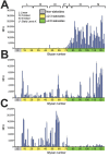
Receptor-binding specificities of influenza A viruses, New York, NY, USA. A) A representative human virus, A/Kawasaki/ 173-PR8(H1N1) is shown for comparison with B) the avian influenza A(H7N2) virus A/chicken/NY/99 and C) the feline influenza A(H7N2) virus A/feline/NY/16. Receptor-binding specificities of the avian and feline viruses were compared with those of the human virus in a glycan microarray containing α2,3- and α2,6-linked sialosides. Error bars represent SDs calculated from 4 replicate spots of each glycan. RFU, relative fluorescence units. A complete list of the glycans used is shown in Technical Appendix Table 2.
Figure 7
Figure 7
Distribution of α2,3- and α2,6-linked sialosides in the respiratory organs of a cat, New York, NY, USA. The α2,3- and α2,6-linked sialosides in the respiratory organs of a naïve cat were detected with biotinylated Maackia amurensis lectin I or II (MAA I, MAA II) or Sambucus nigra lectin (SNA I), respectively. Inset shows closer view of MAA III binding with alveolar epithelium in the lung. Plus signs (+) indicate that sialosides were detected. Scale bars indicate 50 μm.

References

    1. Wright PF, Neumann G, Kawaoka Y. Orthomyxoviruses. In: Knipe DM, Howley PM, Cohen JI, Griffin DE, Lamb RA, Martin MA, et al., editors. Fields Virology. Sixth ed. Philadelphia, PA, USA: Lippincott Williams & Wilkins; 2013. p. 1186–243.
    1. Crawford PC, Dubovi EJ, Castleman WL, Stephenson I, Gibbs EP, Chen L, et al. Transmission of equine influenza virus to dogs. Science. 2005;310:482–5. 10.1126/science.1117950 - DOI - PubMed
    1. Xie X, Ma K, Liu Y. Influenza A virus infection in dogs: epizootiology, evolution and prevention - A review. Acta Vet Hung. 2016;64:125–39. 10.1556/004.2016.014 - DOI - PubMed
    1. Song D, Kang B, Lee C, Jung K, Ha G, Kang D, et al. Transmission of avian influenza virus (H3N2) to dogs. Emerg Infect Dis. 2008;14:741–6. 10.3201/eid1405.071471 - DOI - PMC - PubMed
    1. Lee YN, Lee DH, Lee HJ, Park JK, Yuk SS, Sung HJ, et al. Evidence of H3N2 canine influenza virus infection before 2007. Vet Rec. 2012;171:477. 10.1136/vr.100718 - DOI - PubMed

Publication types

MeSH terms

LinkOut - more resources