. 2018 Feb 6;57(5):772-780.
doi: 10.1021/acs.biochem.7b01083. Epub 2018 Jan 5.

Redox-Inactive Peptide Disrupting Trx1-Ask1 Interaction for Selective Activation of Stress Signaling

Affiliations

Redox-Inactive Peptide Disrupting Trx1-Ask1 Interaction for Selective Activation of Stress Signaling

Dilini N Kekulandara et al. Biochemistry. .

Abstract

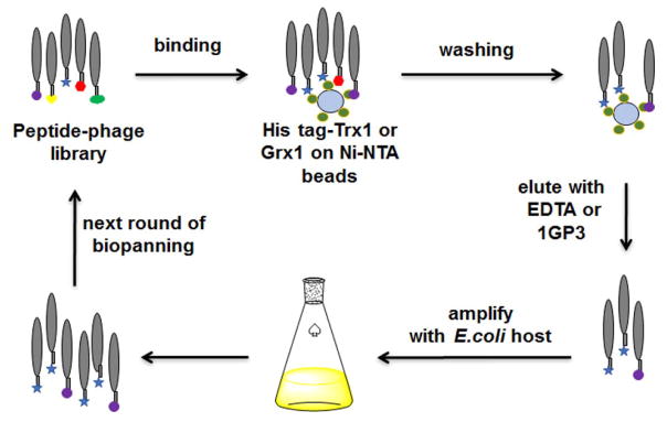
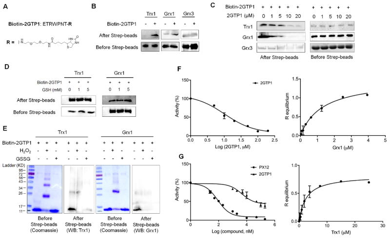
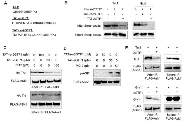
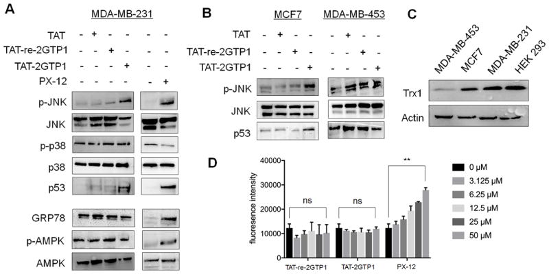
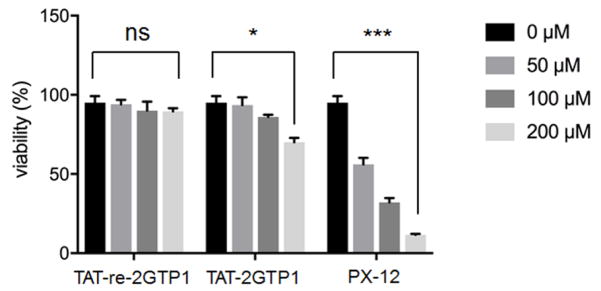
Thioredoxin 1 (Trx1) and glutaredoxin 1 (Grx1) are two ubiquitous redox enzymes that are central for redox homeostasis but also are implicated in many other processes, including stress sensing, inflammation, and apoptosis. In addition to their enzymatic redox activity, a growing body of evidence shows that Trx1 and Grx1 play regulatory roles via protein-protein interactions with specific proteins, including Ask1. The currently available inhibitors of Trx1 and Grx1 are thiol-reactive electrophiles or disulfides that may suffer from low selectivity because of their thiol reactivity. In this report, we used a phage peptide library to identify a 7-mer peptide, 2GTP1, that binds to both Trx1 and Grx1. We further showed that a cell-permeable derivative of 2GTP1, TAT-2GTP1, disrupts the Trx1-Ask1 interaction, which induces Ask1 phosphorylation with subsequent activation of JNK, stabilization of p53, and reduced viability of cancer cells. Notably, as opposed to a disulfide-derived Trx1 inhibitor (PX-12), TAT-2GTP1 was selective for activating the Ask1 pathway without affecting other stress signaling pathways, such as endoplasmic reticulum stress and AMPK activation. Overall, 2GTP1 will serve as a useful probe for investigating protein interactions of Trx1.


Conflict of interest statement

Notes

The authors declare no competing financial interest.

Figures

Figure 1
Figure 1
Identification of Trx1- or Grx1-binding peptides via phage library screening. Bound phage were eluted by EDTA or 1GP3.
Figure 2
Figure 2
In vitro binding and inhibitory analyses of 2GTP1 with Trx1 and Grx1. (A) The structure of biotinylated 2GTP1 (bitotin-2GTP1). (B–E) The binding analyses of 2GTP1 with Trx1 and Grx1. Biotin-2GTP1 was immobilized on streptavidin beads, and incubated with purified Trx1, Grx1, or Grx3 (B), which was repeated in the presence of non-biotinylated 2GTP1 as a competitor (C), reduced glutathione (D), or oxidants (10 molar equivalents to Trx1 or Grx1) (E). Trx1, Grx1, or Grx3 was analyzed by Western blotting before and after streptavidin beads. All Western blots represent data from two or three independent experiments. (F) Inhibitory activity of 2GTP1 for Grx1 deglutathionylation (left) and the binding affinity of 2GTP1 to Grx1 measured by BLI (right). (G) Inhibitory activity of 2GTP1 for Trx1-mediated insulin reduction (left) and the binding affinity of 2GTP1 to Trx1 measured by BLI (right). Data represent the mean ± SD, n= 3 independent experiments.
Figure 3
Figure 3
A cell-permeable derivative of 2GTP1 (TAT-2GTP1) for disruption of the Ask1-Trx1 interaction and activation of Ask1. (A) The sequences of TAT, TAT-2GTP1, and TAT-re-2GTP1 that contains a reverse sequence of 2GTP1. (B) In vitro validation of TAT-2GTP1 and TAT-re-2GTP1 for binding to Trx1 (left) or Grx1 (right). TAT-2GTP1 or TAT-re-2GTP1 was added to a solution containing Trx1 or Grx1 bound to biotin-2GTP1 on streptavidin beads. Bound Trx1 or Grx1 was analyzed by Western blotting. (C) Disruption of the Ask1-Trx1 interaction by TAT-2GTP1. Ask1-Trx1 interaction was analyzed by co-IP after incubation of TAT-2GTP1 to MDA-MB-231 cells for 6 h. (D) Ask1 activation by TAT-2GTP1. Ask1 phosphorylation was analyzed after incubation of TAT-2GTP1 to MDA-MB-231 cells for 6 h. (E) Disruption of Ask1-Trx1 and Ask1-Grx1 interaction by 2GTP1 in vitro. The transfected FLAG-Ask1 was pull-downed by FLAG-antibody, and incubated with purified Trx1 (top) or Grx1 (bottom) in vitro in the presence of 2GTP1. Bound Trx1 or Grx1 was analyzed by Western blotting. All Western blots represent data from two or three independent experiments.
Figure 4
Figure 4
The evaluation of TAT-2GTP1 on ROS induction and stress signaling pathways. (A–B) The effect of TAT-2GTP1 versus PX-12 on Ask1 downstream signaling pathway, ER stress, or AMPK activation in MDA-MB-231 (A), MCF7, and MDA-MB-453 cell lines (B). TAT-2GTP1, TAT-re-2GTP1, TAT, or PX-12 (all 100 μM) was incubated for 6 h. Lysates were analyzed by Western blotting. (C) The level of Trx1 in different cell lines. All Western blots represent data from two or three independent experiments. (D) The level of ROS induced by TAT-2GTP1 versus PX-12. After incubation of TAT-2GTP1, TAT-re-2GTP1, or PX-12 to MDA-MB-231 cells for 6 h, a DCF-DA assay was used to measure induction of ROS. Data represent the mean ± SD, n= 2 independent experiments. Difference is significant by two-tailed Student’s unpaired t-test with Welch’s correction, *p < 0.05, **p < 0.01, ***p < 0.001, and ns (not significant, p > 0.05).
Figure 5
Figure 5
Cell viability induced by TAT-2GTP1. Trypan blue assay was used to measure viability of MDA-MB-231 cells after incubation of TAT-2GTP1, TAT-re-2GTP1 or PX-12 for 48 h. Data represent the mean ± SD, n= 2 independent experiments. Difference is significant by two-tailed Student’s unpaired t-test with Welch’s correction, *p < 0.05, **p < 0.01, ***p < 0.001, and ns (not significant, p > 0.05).

References

    1. Meyer Y, Buchanan BB, Vignols F, Reichheld JP. Thioredoxins and Glutaredoxins: Unifying Elements in Redox Biology. Annu Rev Genet. 2009;43:335–367. - PubMed
    1. Hanschmann EM, Godoy JR, Berndt C, Hudemann C, Lillig CH. Thioredoxins, Glutaredoxins, and Peroxiredoxins-Molecular Mechanisms and Health Significance: from Cofactors to Antioxidants to Redox Signaling. Antioxid Redox Sign. 2013;19:1539–1605. - PMC - PubMed
    1. Velu CS, Niture SK, Doneanu CE, Pattabiraman N, Srivenugopal KS. Human p53 is inhibited by glutathionylation of cysteines present in the proximal DNA-Binding domain during oxidative stress. Biochemistry-Us. 2007;46:7765–7780. - PMC - PubMed
    1. Ueno M, Masutani H, Arai RJ, Yamauchi A, Hirota K, Sakai T, Inamoto T, Yamaoka Y, Yodoi J, Nikaido T. Thioredoxin-dependent redox regulation of p53-mediated p21 activation. J Biol Chem. 1999;274:35809–35815. - PubMed
    1. Reynaert NL, van der Vliet A, Guala AS, McGovern T, Hristova M, Pantano C, Heintz NH, Heim J, Ho YS, Matthews DE, Wouters EFM, Janssen-Heininger YMW. Dynamic redox control of NF-kappa B through glutaredoxin-regulated S-glutathionylation of inhibitory kappa B kinase beta. P Natl Acad Sci USA. 2006;103:13086–13091. - PMC - PubMed

Publication types

MeSH terms