

Age-related changes in baroreflex sensitivity and cardiac autonomic tone in children mirrored by regional brain gray matter volume trajectories
- PMID: 29261644
- PMCID: PMC5866169
- DOI: 10.1038/pr.2017.273
Age-related changes in baroreflex sensitivity and cardiac autonomic tone in children mirrored by regional brain gray matter volume trajectories
Abstract
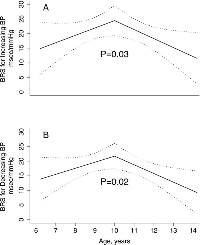
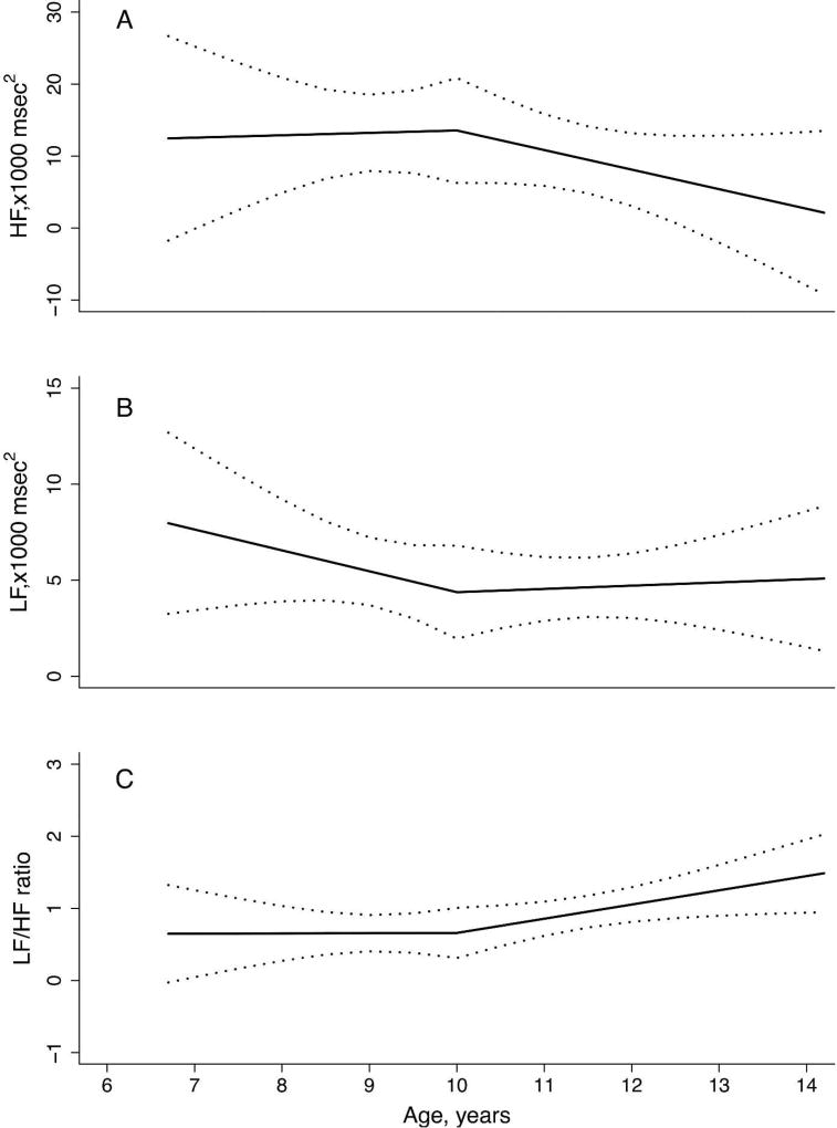
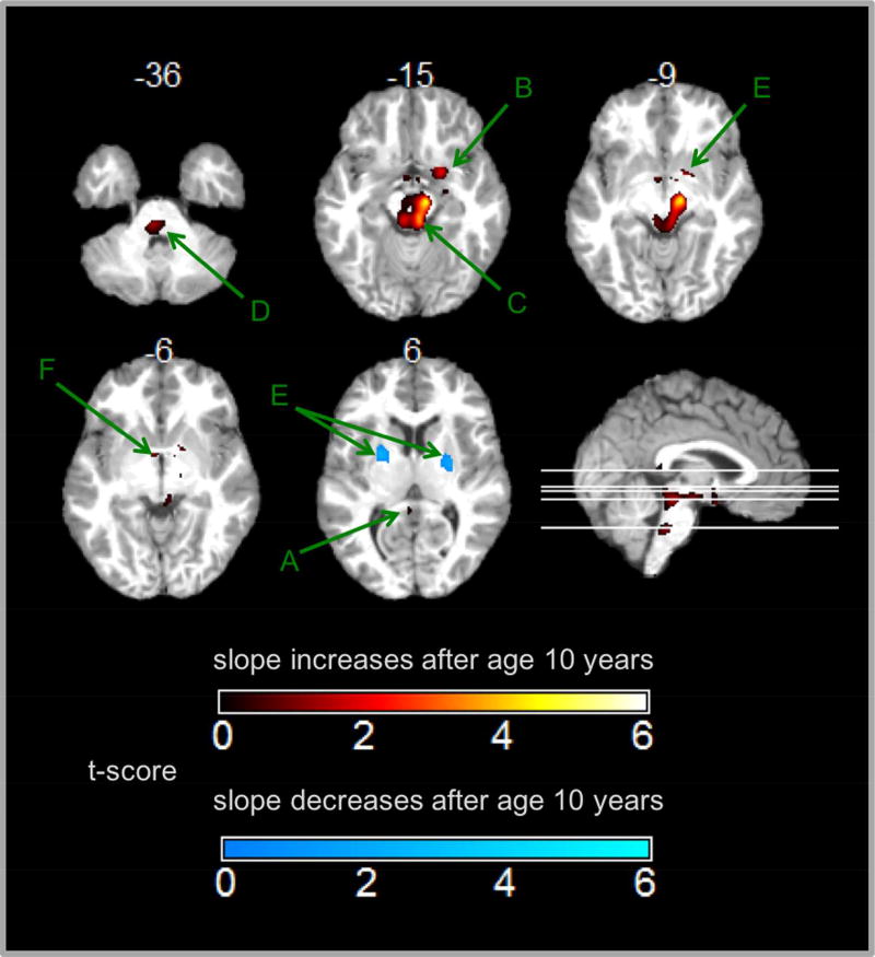
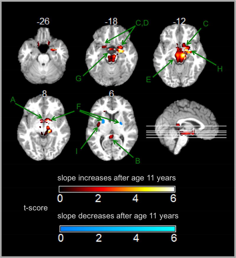
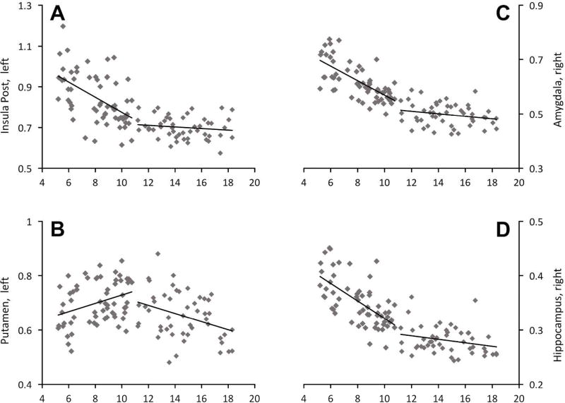
BackgroundThe baroreflex and central autonomic brain regions together control the cardiovascular system. Baroreflex sensitivity (BRS) decreases with age in adults. Age-related changes in brain regions for cardiovascular control in children are unknown. We studied age-related changes in BRS, cardiac autonomic tone, and gray matter volume (GMV) of brain regions associated with cardiovascular control.MethodsBeat-to-beat blood pressure and heart rate (HR) were recorded in 49 children (6-14 years old). Spontaneous BRS was calculated by the sequence method. Cardiac autonomic tone was measured by spectral analysis of HR variability. GMV was measured using voxel-based morphometryin 112 healthy children (5-18 years old).ResultsAge-related changes in BRS were significantly different in children <10 years and ≥10 years. Age-related changes in GMV in regions of interest (ROI) were also significantly different between children <10 and ≥10 years and between children <11 and ≥11 years. However, age-related changes in cardiac autonomic tone were progressive.ConclusionsSignificant changes in BRS trajectories between <10 and ≥10 years may be associated with similar age-related changes of GMV in brain ROI. This new knowledge will guide future studies examining whether childhood cardiovascular disruption manifests as deviated maturation trajectories of specific brain regions.
Figures
Similar articles
-
Central autonomic network functional connectivity: correlation with baroreflex function and cardiovascular variability in older adults.Brain Struct Funct. 2020 Jun;225(5):1575-1585. doi: 10.1007/s00429-020-02075-w. Epub 2020 Apr 30. Brain Struct Funct. 2020. PMID: 32350644 Free PMC article.
-
Trigonometric regressive spectral analysis reliably maps dynamic changes in baroreflex sensitivity and autonomic tone: the effect of gender and age.PLoS One. 2010 Aug 16;5(8):e12187. doi: 10.1371/journal.pone.0012187. PLoS One. 2010. PMID: 20808439 Free PMC article.
-
Measures of cardiovascular autonomic regulation derived from spontaneous methods and the Valsalva maneuver.Auton Neurosci. 2003 Jan 31;103(1-2):100-5. doi: 10.1016/s1566-0702(02)00151-0. Auton Neurosci. 2003. PMID: 12531403
-
A review of human neuroimaging investigations involved with central autonomic regulation of baroreflex-mediated cardiovascular control.Auton Neurosci. 2017 Nov;207:10-21. doi: 10.1016/j.autneu.2017.05.008. Epub 2017 May 15. Auton Neurosci. 2017. PMID: 28529059 Review.
-
Baroreflex sensitivity: measurement and clinical implications.Ann Noninvasive Electrocardiol. 2008 Apr;13(2):191-207. doi: 10.1111/j.1542-474X.2008.00219.x. Ann Noninvasive Electrocardiol. 2008. PMID: 18426445 Free PMC article. Review.
Cited by
-
Effects of age and sex on vasomotor activity and baroreflex sensitivity during the sleep-wake cycle.Sci Rep. 2022 Dec 27;12(1):22424. doi: 10.1038/s41598-022-26440-3. Sci Rep. 2022. PMID: 36575245 Free PMC article.
-
Role of Vascular Receptors in the Development of Hypertension in the Elderly Population.Int J Angiol. 2022 Dec 30;31(4):260-266. doi: 10.1055/s-0042-1759650. eCollection 2022 Dec. Int J Angiol. 2022. PMID: 36588863 Free PMC article.
-
Arterial Baroreceptor Physiology: Differences Between Normal Subjects and Pediatric Patients with Postural Tachycardia and Neurocardiogenic Syncope.Pediatr Cardiol. 2022 Jun;43(5):1011-1019. doi: 10.1007/s00246-022-02815-1. Epub 2022 Jan 28. Pediatr Cardiol. 2022. PMID: 35089394
References
-
- Giedd JN, Blumenthal J, Jeffries NO, et al. Brain development during childhood and adolescence: a longitudinal MRI study. Nat Neurosci. 1999;2:861–863. - PubMed
-
- Walhovd KB, Tamnes CK, Fjell AM. Brain structural maturation and the foundations of cognitive behavioral development. Curr Opin Neurol. 2014;27:176–184. - PubMed
Publication types
MeSH terms
Grants and funding
LinkOut - more resources
Full Text Sources
Other Literature Sources
