. 2017 Dec 20;12(12):e0188917.
doi: 10.1371/journal.pone.0188917. eCollection 2017.

Decreased expression of lethal giant larvae causes ovarian follicle cell outgrowth in the Drosophila Scutoid mutant

Affiliations

Decreased expression of lethal giant larvae causes ovarian follicle cell outgrowth in the Drosophila Scutoid mutant

Chen-Yuan Tseng et al. PLoS One. .

Abstract

Snail, a zinc-finger transcription factor, controls the process of epithelial-mesenchymal transition, and ectopic expression of this protein may produce cells with stem cell properties. Because the effect of Snail expression in ovarian epithelial cells remains unclear, we generated Drosophila ovarian follicle stem cells (FSCs) with homozygous Scutoid (Sco) mutation. The Sco mutation is a reciprocal transposition that is known to induce ectopic Snail activity. We found that Sco mutant FSCs showed excess proliferation and high competitiveness for niche occupancy, and the descendants of this lineage formed outgrowths that failed to enter the endocycle. Surprisingly, such phenotypes were not rescued by suppressing Snail expression, but were completely restored by supplying Lethal giant larvae (Lgl). The lgl allele is a cell polarity gene that is often mutated in the genome. Importantly, Sco mutants survived in a complementation test with lgl. This result was probably obtained because the Sco-associated lgl allele appears to diminish, but not ablate lgl expression. While our data do not rule out the possibility that the Sco mutation disrupts a regulator of lgl transcription, our results strongly suggest that the phenotypes we found in Sco mutants are more closely associated with the lgl allele than ectopic Snail activity.


Conflict of interest statement

Competing Interests: The authors have declared that no competing interests exist.

Figures

Fig 1
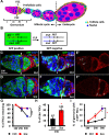
Fig 1. Sco FSCs exhibit extended lifespan, enhanced proliferation, and increased competitiveness for niche occupancy.
(A) Schematic of the Drosophila ovariole. The anterior-most structure of the ovariole, the germarium, contains germ cells that are enveloped by prefollicle cells (light green), to form egg chambers. The prefollicle cells are derived from two FSCs (yellow), which are located at the 2a/2b boundary of the germarium. Follicle cells of egg chambers up to stage (S) 6 (light blue) undergo mitotic cycles, while follicle cells of egg chambers after stage 7 (orange) enter the endocycle. (B) Mitotic recombination was used to generate Sco FSCs. Females were generated, carrying a wild-type allele linked to a marker gene (GFP) in trans with the Sco allele. FLP-mediated recombination between FRT sites during mitotic division generated a homozygous Sco FSC that could be identified by the absence of GFP. (C-F) Control (Ctrl) (C and E) and Sco mosaic germaria (D and F) shown at one week (W) after clone induction (ACI): GFP (green, wild-type cells), FasIII (red, follicle cell lineages), DAPI (blue, DNA), and Edu (white in E and F, indicating proliferating cells). Solid and empty triangles indicate GFP-positive and GFP-negative FSCs, respectively. Scale bar, 10 μm. (G) Relative percentage (%) of germaria carrying GFP-negative follicle cell clones at 1, 2 and 3 weeks ACI. (H) Percentage of Edu-positive FSC clones in total FSC clones at one week ACI. The number of FSC clones analyzed is shown above the bar. (I) Percentage of germaria carrying two GFP-negative FSCs in control (black line) and Sco (red line) at 1, 2 and 3 weeks ACI. The blue squares in G and I indicate significant differences as compared to the initial time point. * P<0.05. *** P<0.001. Statistical analysis was carried out with student t-test. Data are shown as mean ± SEM. The genotype of C and E is hs-flp/+; ubi-gfpFRT40A/FRT40A, of D and F is hs-flp/+; ubi-gfpFRT40A/ScoFRT40A.
Fig 2
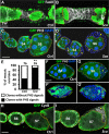
Fig 2. Sco follicle cells are hyperproliferative and do not enter the endocycle.
Control (Ctrl) (A, C, F and H) and Sco mosaic ovarioles (B, D, G and I) at one week (1W) after clone induction (ACI) are labeled with GFP (green, wild-type cells) and FasIII (gray, membranes of follicle cell lineages) in A and B, phospho-Histone 3 (PH3, gray, mitotic marker) and DAPI (blue, DNA) in C, D, F and G, and Cyclin B (CycB, gray, G2/M phase marker) in H and I. Wild-type cells are outlined by yellow dashed lines. The scale bar in A is 10 μm, and scale bars in C, F and H are 20 μm. (A and B) Sco mosaic ovarioles contain stalk cell overgrowths that are completely composed of excessive numbers of irregularly-shaped Sco cells, as compared to the control. (C and D) Sco follicle cells formed multiple layers in stage (S) 4 and 6 egg chambers. (E) Percentage (%) of mosaic ovarioles exhibiting PH3 signal in GFP-negative follicle cell clones. The number of ovarioles analyzed is shown above each bar. (F and G) Sco follicle cells of the stage 8 egg chamber formed multiple layers, and continued to undergo mitosis. F’ and G’ show the outermost layers of egg chambers. Asterisks indicate nurse cells. (H and I) Sco follicle cells of the stage 7 egg chamber retained greater CycB signals than the controls. Wild-type cells are outlined by yellow dashed lines. Asterisks indicate anterior or posterior poles of egg chambers. The genotype of the controls in A, C, E, F and H is hs-flp/+; ubi-gfpFRT40A/ FRT40A, and of the Sco mosaic mutant in B, D, E, G and I is hs-flp/+; ubi-gfpFRT40A/ScoFRT40A.
Fig 3
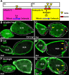
Fig 3. Sco follicle cells lose cell polarity and are delaminated.
(A-F) One-week (w)-old control (ctrl) (A, C and E) and Sco mosaic (B, D and F) stage (S) 6 and 7 egg chambers: GFP (green, wild-type cells) and E-cadherin (gray, E-cad) in A and B; Disc large (gray, Dlg) in C and D; aPKC (E and F); DAPI (blue, DNA). A’ and B’ show E-cad channel only; C’ and D’ show Dlg channel only; E’ and F’ show aPKC only. Dashed lines indicate follicle cell clones. Inserts are enlarged images from the area indicated by asterisks. Scale bar, 20 μm. The genotype of A, C and E is hs-flp/+; ubigfp FRT40A/ FRT40A, of B, D and F is hs-flp/+; ubi-gfpFRT40A/ScoFRT40A.
Fig 4
Fig 4. Notch signaling is disturbed in the outer layers of Sco follicle cells.
(A) Notch signaling is required for the transition of follicle cells from the mitotic phase to endocycle phase [10]. In mitotic follicle cells (up to stage 6), Notch signaling activity is low because germ cells produce low amounts of Delta (a Notch ligand, shown as yellow triangles). In addition, Cut is expressed in follicle cells to suppress the mitosis-endocytosis transition. After stage 7, Notch signaling is activated to promote Hindsight expression, which suppresses Cut expression and thereby permits the mitosis-endocytosis transition. Red squares indicate Notch receptors. (B-G) One-week (w)-old control (ctrl) (B, D and F) and Sco mutant mosaic egg chambers (C, E and G) at stages (S) 6 and 7: GFP (green, wild-type cells), Esplm7-lacZ (gray, a Notch signaling reporter) in B and C, Cut (gray) in D and E, Hindsight (gray) in F and G. Expression of E(spl)m7-lacZ is decreased in ectopic layers of Sco mutant follicle cells located far from the germline at the anterior and posterior poles. In the control, Cut is mainly expressed in stage 6 follicle cells and is downregulated in stage 7 follicle cells. However, Cut expression is weaker in the inner layer as compared to the outer layer of Sco mutant follicle cells. In contrast, Hindsight expression is stronger in the inner layer as compared to the outer layer of Sco follicle cells at stage 7, indicating a non-cell autonomous effect of Sco on the mitosis-endocycle transition. Arrows show the boundary between germ cells and follicle cells. Asterisks indicate outer layers of ectopic follicle cells. Scale bar, 20 μm. The genotype of B, D and F is hs-flp/+; ubi-gfpFRT40A/FRT40A, of C, E and G is hs-flp/+; ubi-gfpFRT40A/ScoFRT40A.
Fig 5
Fig 5. Snail and Noc do not account for the multiple-layered phenotype of Sco follicle cells.
(A-D) One-week-old control (ctrl) mosaic (A), Sco mosaic (B) and Sco mosaic ovarioles with snail knockdown (C and D). (E-J) One-week (W)-old control (ctrl) (E and F), 10930>nocRNAi (G), GR1> nocRNAi (H), 10930>snail & gfp (I), and 10930>snail & nocRNAi ovarioles (J): FasIII (green, follicle cell lineages), Traffic Jam (Tj) (gray, follicle cells) in I and J, and DAPI (blue, DNA). Arrows in I and J indicated overexpression of Snail increase cell number in the stalk that connects two egg chambers. The scale bar is 20 μm. The genotype of A is c587-GAL4/UAS-flp; ubi-gfpFRT40A/FRT40A, of B, C, and D is c587-GAL4/UAS-flp: ScoFRT40A/ubi-gfpFRT40A.
Fig 6
Fig 6. Exogenous supplement of lgl prevents Sco follicle cells forming multiple layers.
(A) Mosaic Analysis with Repressible Cell Marker (MARCM) was used to generate Sco homozygous mutant cells expressing lgl-gfp. Females carried wild-type alleles linked to tubulin promoter-GAL80 (a GAL4 suppressor) in trans with the Sco mutant allele on the second chromosome, actin promoter-GAl4, UAS-nuclear (n) LacZ and UAS-lgl-gfp on the third chromosome. FLP-mediated recombination between two FRT sites during mitotic division generated Sco homozygous mutant cells lacking GAL80, allowing UAS-trangenes to be expressed by GAL4 driven by an actin promoter. (B-C) Mosaic egg chambers in one-week (W)-old Sco mutant (B), and Sco mutant with lgl overexpression: LacZ (green, mutant cells) and DAPI (blue, DNA). Dashed lines mark follicle cell clones. The scale bar is 20 μm. The genotype of B is hs-flp/+; tub-Gal80FRT40A/ ScoFRT40A; act-GAL4UAS-nlacZ/+, and of C is hs-flp/+; tub-Gal80FRT40A/ ScoFRT40A; act-GAL4UAS-nlacZ/UAS-lgl-gfp.

Similar articles

References

    1. Kalluri R, Weinberg RA. The basics of epithelial-mesenchymal transition. J Clin Invest. 2009;119(6):1420–8. doi: 10.1172/JCI39104 ; PubMed Central PMCID: PMCPMC2689101. - DOI - PMC - PubMed
    1. Cano A, Perez-Moreno MA, Rodrigo I, Locascio A, Blanco MJ, del Barrio MG, et al. The transcription factor snail controls epithelial-mesenchymal transitions by repressing E-cadherin expression. Nat Cell Biol. 2000;2(2):76–83. doi: 10.1038/35000025 . - DOI - PubMed
    1. Wang H, Wang HS, Zhou BH, Li CL, Zhang F, Wang XF, et al. Epithelial-mesenchymal transition (EMT) induced by TNF-alpha requires AKT/GSK-3beta-mediated stabilization of snail in colorectal cancer. PLoS One. 2013;8(2):e56664 doi: 10.1371/journal.pone.0056664 ; PubMed Central PMCID: PMCPMC3576347. - DOI - PMC - PubMed
    1. Masui T, Ota I, Yook JI, Mikami S, Yane K, Yamanaka T, et al. Snail-induced epithelial-mesenchymal transition promotes cancer stem cell-like phenotype in head and neck cancer cells. Int J Oncol. 2014;44(3):693–9. doi: 10.3892/ijo.2013.2225 . - DOI - PubMed
    1. Zhou W, Lv R, Qi W, Wu D, Xu Y, Liu W, et al. Snail contributes to the maintenance of stem cell-like phenotype cells in human pancreatic cancer. PLoS One. 2014;9(1):e87409 doi: 10.1371/journal.pone.0087409 ; PubMed Central PMCID: PMCPMC3906155. - DOI - PMC - PubMed

Substances