. 2017 Dec 19;12(12):e0189629.
doi: 10.1371/journal.pone.0189629. eCollection 2017.

A mutant of phosphomannomutase1 retains full enzymatic activity, but is not activated by IMP: Possible implications for the disease PMM2-CDG

Affiliations

A mutant of phosphomannomutase1 retains full enzymatic activity, but is not activated by IMP: Possible implications for the disease PMM2-CDG

Valentina Citro et al. PLoS One. .

Abstract

The most frequent disorder of glycosylation, PMM2-CDG, is caused by a deficiency of phosphomannomutase activity. In humans two paralogous enzymes exist, both of them require mannose 1,6-bis-phosphate or glucose 1,6-bis-phosphate as activators, but only phospho-mannomutase1 hydrolyzes bis-phosphate hexoses. Mutations in the gene encoding phosphomannomutase2 are responsible for PMM2-CDG. Although not directly causative of the disease, the role of the paralogous enzyme in the disease should be clarified. Phosphomannomutase1 could have a beneficial effect, contributing to mannose 6-phosphate isomerization, or a detrimental effect, hydrolyzing the bis-phosphate hexose activator. A pivotal role in regulating mannose-1phosphate production and ultimately protein glycosylation might be played by inosine monophosphate that enhances the phosphatase activity of phosphomannomutase1. In this paper we analyzed human phosphomannomutases by conventional enzymatic assays as well as by novel techniques such as 31P-NMR and thermal shift assay. We characterized a triple mutant of phospomannomutase1 that retains mutase and phosphatase activity, but is unable to bind inosine monophosphate.


Conflict of interest statement

Competing Interests: The authors have declared that no competing interests exist.

Figures

Fig 1
Fig 1. Phosphomannomutase activity of PMM1 and PMM2 on Man-6-P monitored by 31P-NMR spectroscopy.
PMM1 or PMM2 (54 μg) were incubated with Man-6-P 1mM and Glc-1,6-P2 0.1 mM at 27°C. Panel A) Spectrum of reagents prior to PMM1 addition. Panel B) Spectrum of products after PMM1 addition. Panel C) Spectrum of reagents prior to PMM2 addition. Panel D) Spectrum of products after PMM2 addition. The spectra were accumulated over 40 min. Creatine phosphate (1 mM) was added as an internal standard and all the spectra were referred to it (0 ppm). Resonance assignment was obtained by comparison with pure compounds: 1, Glc-6-P; 2, Man-6-P; 3 and 5, Man-1,6-P2; 4, Man-1-P; 6 and 7, Glc-1,6-P2; *, inorganic phosphate.
Fig 2
Fig 2. Phosphomannomutase activity of PMM1 and PMM2 on Man-1-P monitored by 31P-NMR spectroscopy.
PMM1 or PMM2 (54 μg) were incubated with Man-1-P 0.5 mM and Glc-1,6-P2 0.1 mM at 27°C, Panel A) Spectrum of reagents prior to PMM1 addition. Panel B) Spectrum of products after PMM1 addition. Panel C) Spectrum of reagents prior to PMM2 addition. Panel D) Spectrum of products after PMM2 addition. The spectra were accumulated over 40 min. Creatine phosphate (1 mM) was added as an internal standard and all the spectra were referred to it (0 ppm). Resonance assignment was obtained by comparison with pure compounds: 1, Glc-6-P; 2, Man-6-P; 3 and 5, Man-1,6-P2; 4, Man-1-P; 6 and 7, Glc-1,6-P2; *, inorganic phosphate; °, unidentified peaks.
Fig 3
Fig 3. Phosphatase activity of PMM1 monitored by 31P-NMR spectroscopy.
PMM1 (20 μg) was incubated with Glc-1,6-P2 0.55 mM at 27°C, with and without IMP 0.17 mM. Panel A) Spectrum of reagents, Glc-1,6-P2 and IMP, prior to PMM1 addition. Panel B) Spectrum of products and IMP after PMM1 addition. Panel C) Spectrum of reagents, Glc-1,6-P2, prior to PMM1 addition. Panel D) Spectrum of products after PMM1 addition. Creatine phosphate (1 mM) was added as an internal standard and all the spectra were referred to it (0 ppm). Resonance assignment was obtained by comparison with pure compounds: 1 and 2, Glc-1,6-P2; 3 Glc-1-P+Pi; 4, Glc-6-P; 5, IMP; *, inorganic phosphate.
Fig 4
Fig 4. PMM1 and PMM2 sequence alignment.
Active site residues are highlighted in grey, residues that are conserved in PMM1 orthologous proteins, but not in PMM2 orthologous proteins are in bold.
Fig 5
Fig 5. Effects of IMP on the phosphatase activity of PMM1, QDK-PMM1 and PMM2.
Panel A) The substrate was Man-1,6-P2 0.08 mM with or without IMP 0.17 mM. Panel B) The substrate was Glc-1,6-P2 0.145 mM with or without IMP 0.17 mM.
Fig 6
Fig 6. Competitive inhibition of PMM1 mutase activity by IMP.
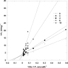
Phosphoglucomutase activity of PMM1 (Glc-1,6-P2 1 μM and Glc-1-P ranging from 0 to 60 μM) was measured at 0, 2, 4, 6 and 10 μM IMP. Data are shown as Lineweaver-Burk plots.
Fig 7
Fig 7. Effects of bisphosphonates on the phosphatase and glucomutase activities of PMM1, QDK-PMM1 and PMM2.
Panel A) Phosphoglucomutase activity of PMM1 and PMM2 was measured in the presence of Glc-1-P 40 μM and Glc-1,6-P2 27 μM. Panel B) Glc-1,6-P2-phosphatase activity of PMM1, QDK-PMM1 and PMM2 was measured in the presence of Glc-1,6-P2 0.145 mM. In both cases the activities were also measured in the presence of clodronate (2.8 mM) or neridronate (1.5 mM).
Fig 8
Fig 8. Thermal stability of phosphomannomutases.
Panel A) Melting temperatures of PMM1 and QDK-PMM1 (0.5 mg/ml) were measured in the presence of different ligands: Glc-1-P 0.5 mM, Glc-1-P 0.5 mM + vanadate 0.5 mM, vanadate 0.5 mM, Glc-1,6-P2 0.5 mM, IMP 0.17 mM. Panel B). Melting temperatures of PMM1 and PMM2 (0.5 mg/ml) were measured in the presence of different concentrations of Glc-1,6-P2 (ranging from 0 to 1 mM). The experiments were conducted at pH 7.5 in the presence of 2.4x SyproOrange by Thermal Shift Assay and the temperature increase was 1°/min from 20 to 90°C.
Fig 9
Fig 9. In silico docking of IMP and PMM1.
Panel A) Unconstrained simulation. Only 5 trajectories are shown. Structure 52, used for the next refinement simulation, is identified by an asterisk. Panel B) Refinement simulation for structure 52 from traj1 in panel A. The circle, the section mark and the asterisk represent the structure further analyzed (see Fig 10 panel B).
Fig 10
Fig 10. Ligand binding interactions.
Panel A) A zoom-in of the interaction between IMP and PMM1 as it is seen in the low energy structure 52 (see Fig 7, panel A): the cap domain is shadowed in light magenta, the core domain is shadowed in grey. Panel B) Refined binding modes: details from energetic minima structures from 3 different trajectories (asterisk -traj5, section mark -traj13, circle -traj14 in Fig 9, panel B).

Similar articles

Cited by

References

    1. Matthijs G, Schollen E, Pardon E, Veiga-Da-Cunha M, Jaeken J, Cassiman JJ, et al. Mutations in PMM2, a phosphomannomutase gene on chromosome 16p13, in carbohydrate-deficient glycoprotein type I syndrome (Jaeken syndrome). Nat Genet. 1997;16(1):88–92. doi: 10.1038/ng0597-88 . - DOI - PubMed
    1. Jaeken J, Hennet T, Freeze HH, Matthijs G. On the nomenclature of congenital disorders of glycosylation (CDG). J Inherit Metab Dis. 2008;31(6):669–72. Epub 2008/10/25. doi: 10.1007/s10545-008-0983-x . - DOI - PubMed
    1. Aebi M. N-linked protein glycosylation in the ER. Biochim Biophys Acta. 2013;1833(11):2430–7. Epub 2013/04/16. doi: 10.1016/j.bbamcr.2013.04.001 [pii]. . - DOI - PubMed
    1. Maeda Y, Kinoshita T. Dolichol-phosphate mannose synthase: structure, function and regulation. Biochim Biophys Acta. 2008;1780(6):861–8. Epub 2008/04/05. doi: 10.1016/j.bbagen.2008.03.005 [pii]. . - DOI - PubMed
    1. Pirard M, Collet JF, Matthijs G, Van Schaftingen E. Comparison of PMM1 with the phosphomannomutases expressed in rat liver and in human cells. FEBS Lett. 1997;411(2–3):251–4. . - PubMed

MeSH terms

Supplementary concepts