

Early detection of Mycobacterium avium subsp. paratuberculosis infection in cattle with multiplex-bead based immunoassays
- PMID: 29261761
- PMCID: PMC5736219
- DOI: 10.1371/journal.pone.0189783
Early detection of Mycobacterium avium subsp. paratuberculosis infection in cattle with multiplex-bead based immunoassays
Abstract
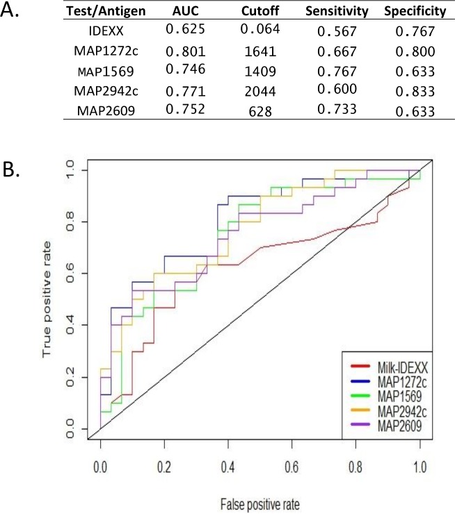
Johne's Disease (JD), caused by Mycobacterium avium subspecies paratuberculosis (MAP), results in significant economic loss to livestock production. The early detection of MAP infection in animals with extant serological assays has remained challenging due to the low sensitivity of commercially available ELISA tests, a fact that has hampered the development of effective JD control programs. Our recent protein microarray-based studies identified several promising candidate antigens that are immunogenic during different stages of MAP infection. To evaluate these antigens for use in diagnostic assays and reliably identify animals with MAP infection, a multiplex (Luminex®) assay was developed using color-coded flourescent beads coupled to 6 MAP recombinant proteins and applied to screen 180 serum and 90 milk samples from cows at different stages of MAP infection including negative (NL), fecal test positive/ELISA negative (F+E-), and fecal positive/ELISA positive (F+E+). The results show that while serum antibody reactivities to each of the 6 antigens were highest in F+E+ group, antibody reactivity to three of the six antigens were identified in the F+E- group, suggesting that these three antigens are expressed and provoke antibody responses during the early infection stages with MAP. Further, antibodies against all six antigens were elevated in milk samples from both the F+E- and F+E+ groups in comparison to the NL group (p<0.01). Taken together, the results of our investigation suggest that multiplex bead-based assays are able to reliably identify MAP infection, even during early stages when antibody responses in animals are undetectable with widely used commercial ELISA tests.
Conflict of interest statement
Figures


References
-
- Clarke CJ (1997) The pathology and pathogenesis of paratuberculosis in ruminants and other species. J Comp Pathol 116: 217–261. - PubMed
-
- Stewart DJ, Vaughan JA, Stiles PL, Noske PJ, Tizard ML, Prowse SJ, et al. (2004) A long-term study in Merino sheep experimentally infected with Mycobacterium avium subsp. paratuberculosis: clinical disease, faecal culture and immunological studies. Vet Microbiol 104: 165–178. doi: 10.1016/j.vetmic.2004.09.007 - DOI - PubMed
-
- Ott SL, Wells SJ, Wagner BA (1999) Herd-level economic losses associated with Johne's disease on US dairy operations. Prev Vet Med 40: 179–192. - PubMed
-
- Collins MT, Wells SJ, Petrini KR, Collins JE, Schultz RD, Whitlock RH (2005) Evaluation of five antibody detection tests for diagnosis of bovine paratuberculosis. Clin Diagn Lab Immunol 12: 685–692. doi: 10.1128/CDLI.12.6.685-692.2005 - DOI - PMC - PubMed
MeSH terms
Substances
LinkOut - more resources
Full Text Sources
Other Literature Sources
