Review
. 2018 May:185:99-121.
doi: 10.1016/j.pharmthera.2017.12.004. Epub 2017 Dec 17.

Potential for therapeutic targeting of AKAP signaling complexes in nervous system disorders

Affiliations
Review

Potential for therapeutic targeting of AKAP signaling complexes in nervous system disorders

Angela R Wild et al. Pharmacol Ther. 2018 May.

Abstract

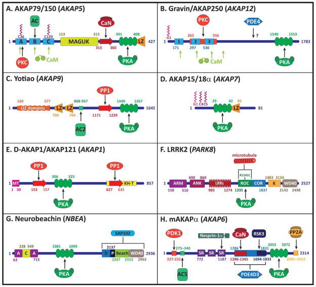
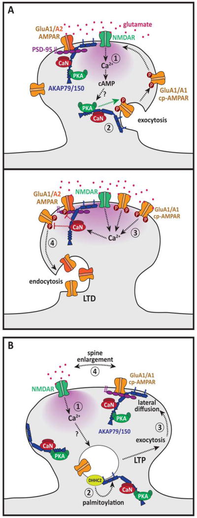
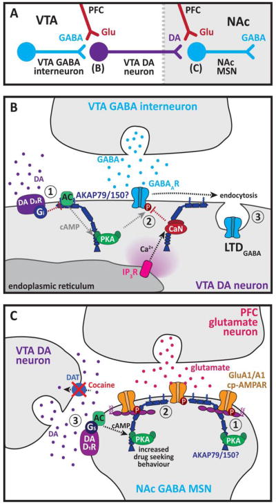
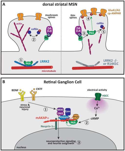
A common feature of neurological and neuropsychiatric disorders is a breakdown in the integrity of intracellular signal transduction pathways. Dysregulation of ion channels and receptors in the cell membrane and the enzymatic mediators that link them to intracellular effectors can lead to synaptic dysfunction and neuronal death. However, therapeutic targeting of these ubiquitous signaling elements can lead to off-target side effects due to their widespread expression in multiple systems of the body. A-kinase anchoring proteins (AKAPs) are multivalent scaffolding proteins that compartmentalize a diverse range of receptor and effector proteins to streamline signaling within nanodomain signalosomes. A number of essential neurological processes are known to critically depend on AKAP-directed signaling and an understanding of the role AKAPs play in nervous system disorders has emerged in recent years. Selective targeting of AKAP protein-protein interactions may be a means to uncouple pathologically active signaling pathways in neurological disorders with a greater degree of specificity. In this review we will discuss the role of AKAPs in both regulating normal nervous system function and dysfunction associated with disease, and the potential for therapeutic targeting of AKAP signaling complexes.

Keywords: AKAP; Calcium; Ion channel; Nervous system; PKA; cAMP.


Conflict of interest statement

11. Conflict of interest statement

The authors declare that there are no conflicts of interest.

Figures

Figure 1
Figure 1. Schematic diagrams of AKAPs discussed in this review
A. AKAP79 (human isoform) anchors PKA, CaN, AC and PKC. Interactions with ion channels occur at the LZ domain and MAGUK binding domain. Membrane targeting is directed by three polybasic domains A, B and C and palmitoylation at Cys-36 and Cys-129. CaM interacts with the three polybasic domains. See main text for references. B. Gravin (human isoform) anchors PKA and PKC (Nauert et al., 1997) and interacts with PDE4 through an unmapped binding site (Willoughby et al., 2006). Membrane targeting is directed by three N-terminal polybasic domains (PB1, 2 and 3) and myristoylation of Gly-1 (Tao et al., 2006; Yan et al., 2009). CaM binds to PB1, 2 and 3, in addition to a fourth CaM binding site designated CB4 (Tao et al., 2006; Schott & Grove, 2013). The central portion of gravin (556–938) associates with the β2-AR (Tao et al., 2003). C. Yotiao anchors PKA, PP1 (Westphal et al., 1999) and AC2 (Piggott et al., 2008). Two LZ motifs and seven leucine/isoleucine zipper (LIZ) motifs regulate interactions with ion channels and IP3Rs (Tu et al., 2004). GluN1 binds between the AC2 and PP1 binding sites (970–1241) (Lin et al., 1998). D. AKAP15/18 (α isoform shown) binds PKA (Fraser et al., 1998) and several ion channels through an LZ motif (Cantrell et al., 1999, 2002; Hulme et al., 2002; Marshall et al., 2011). Membrane targeting is directed by myristoylation and palmitoylation of the N-terminus (Fraser et al., 1998). E. D-AKAP1 anchors PKA (Chen et al., 1997) and PP1 at two binding sites, with the primary PP1 binding site being located toward the C-terminus (Steen et al., 2000; Rogne et al., 2009). The extreme N-terminus contains a mitochondrial targeting domain (Huang et al., 1999). D-AKAP1 is thought to bind mRNAs encoding mitochondrial proteins via the KH and Tudor (KH-T) domains (Ginsberg et al., 2003; Rogne et al., 2006, 2009). See (Merrill & Strack, 2014) for a comprehensive list of binding partners. F. LRRK2 binds PKA within the Ras of complex proteins (ROC) GTPase domain (Parisiadou et al., 2014), which is also the site for microtubule association (Gandhi et al., 2008) and LRRK2 dimerization interactions (Guaitoli et al., 2016). The C-terminal of ROC (COR) domain is thought to act in concert with the ROC domain for GTPase function, while additional enzymatic activity comes from the kinase (KIN) domain. Protein-protein interaction sites include armadillo (ARM), ankyrin (ANK), leucine rich repeats (LRRs) and WD40 domains (Gilsbach & Kortholt, 2014; Cardona et al., 2014). G. Neurobeachin interacts with PKA (Wang et al., 2000) and the synapse-associated protein 102 (SAP102) (Lauks et al., 2012). The N-terminus contains a Concavalin Alike lectin domain (C) domain flanked by two armadillo repeats (A) (Burgess et al., 2009), while the C-terminal is made up of a domain of unknown function 1088 (D) that includes an NLS (Tuand et al., 2016), a pleckstrin homology (P), a beige and Chediak-Higashi (BEACH) and a WD40 domain (Wang et al., 2000; Jogl et al., 2002). H. mAKAPα anchors PKA (Kapiloff et al., 1999), PDK1 (Carlisle Michel et al., 2005), RSK3 (Li et al., 2013a), AC5 (Kapiloff et al., 2009), CaNAβ through a non-PxIxIT motif (Pare et al., 2005a), PDE4D3 (Dodge et al., 2001), PP2A (Dodge-Kafka et al., 2010) and nesprin1α (Pare et al., 2005b). Targeting is achieved by three spectrin repeats (Kapiloff et al., 1999). Shorter isoform mAKAPβ starts at residue 245 (Passariello et al., 2015).
Figure 2
Figure 2. AKAP79/150 and synaptic plasticity
A. AKAP79/150 regulation of LTP and LTD. Top: (1) Both LTP and LTD stimuli activate NMDARs, Ca2+ influx increases cAMP, potentially through activation of adenylyl cyclases 1 and/or 8; (2) AKAP79/150-anchored PKA phosphorylates Ser-845 promoting exocytosis; lateral diffusion and synaptic accumulation of cp-AMAPRs. AKAP79/150 is present in the PSD, extrasynaptic membrane and endosomes and can regulate cp-AMPARs in each of these locations. Bottom: (3) During LTD, Ca2+ influx through cp-AMPARs and NMDARs then activates AKAP79/150-anchored CaN, promoting Ser-845 dephosphorylation; (4) AMPARs are rapidly removed from the synapse along with PSD-95 and AKAP79/150; spine size decreases leading to LTD. Adapted from (Sanderson et al., 2016) B. Dynamic membrane trafficking of AKAP79/150 and LTP. (1) LTP stimulus activates NMDARs; (2) DHHC2 palmitoylates AKAP79/150; (3) AKAP79/150-dependent endosome exocytosis; synaptic delivery of cp-AMPARs and AKAP79/150; (4) spine enlargement and spine LTP.
Figure 3
Figure 3. AKAP79/150 regulation of ion channels and receptors
A. AKAP79/150 interacts with LTCCs through its C-terminal LZ motif (Oliveria et al., 2007). (1) AKAP79/150-anchored PKA regulates LTCC basal phosphorylation at Ser-1700 and Ser-1928, while AKAP79/150-anchored CaN regulates LTCC CDI. (2) Activation of AKAP79/150-anchored CaN by Ca2+ influx through LTCCs leads to dephosphorylation of multiple NFAT phosphorylation sites, uncovering an NLS and promoting NFAT nuclear translocation. NFAT associates with co-transcription factors such as fos and jun. (3) NFAT-dependent transcription increases expression of a number of gene targets including Kv7.2/3 channels. AKAP79/150 also associates with β2ARs and ACs, that enhance PKA activity and phosphorylation of LTCCs. B. Left: Kv4.2 binds to the MAGUK domain of AKAP79/150 (Lin et al., 2011), targeting PKA and CaN to the channel to bi-directionally regulate surface expression and current density. Right: Kv7.2/3 binds to the membrane targeting domains of AKAP79/150 (Hoshi et al., 2003; Bal et al., 2010). (1) Activation of M1Rs within the AKAP79/150 signaling complex promotes activation of PLC; (2) PLC hydrolyses Kv7.2/3 associated PIP2 to IP3 and diacylglycerol (DAG), decreasing channel current; (3) DAG activates AKAP79/150-anchored PKC, which phosphorylates Kv7.2/3, reducing channel affinity for CaM and PIP2. CaM binding to Kv7.2/3 disrupts the Kv7.2/3-AKAP79/150 interaction. C. TRPV1 binds close to the CaN-binding PxIxIT motif of AKAP79/150 (Btesh et al., 2013). Left: PKC phosphorylation of TRPV1 enhances channel sensitivity. Right: Activation of EP4Rs by PGE2 stimulates AKAP79/150-anchored AC and PKA, enhancing TRPV1 sensitivity. AKAP79/150-anchored CaN is engaged during TRPV1 desensitization induced by capsacin. D. Left: AKAP79/150-TRPV1-Kv7.2/3 super-complex. Ca2+ influx through TRPV1 decreases Kv7.2/3 current in AKAP79/150 dependent manner, potentially through promoting PLCδ hydrolysis of PIP2 or enhancing binding of Ca2+-CaM to Kv7.2/3. Right: AKAP79/150-TRPV1-LTCC super-complex. Ca2+ influx through LTCCs desensitizes TRPV1 in an AKAP79/150 dependent manner, potentially through activation of AKAP79/150-anchored CaN.
Figure 4
Figure 4. AKAP regulation of neuronal signalosomes
A. Gravin forms signaling complexes with β2ARs and PKA. (1) β2AR stimulation of AC and gravin-anchored PKA promotes β2AR phosphorylation; (2) β2ARs switch from Gs to Gi coupling; (3) β2AR Gi coupling activates ERK pathway and promotes synaptic potentiation. Adapted from (Havekes et al., 2012). B. Yotiao binds to the GluN1 C-terminal where it regulates NMDAR activity through PKA and PP1 phospho-regulation. Yotiao regulates GABAARs and IP3R1s. AC2 activity is inhibited when anchored to yotiao. C. Top: AKAP15/18α in the hippocampal mossy fiber projection anchors PKA. Pre-synaptic Ca2+ influx activates PKA, promoting pre-synaptic MF-LTP. Bottom: AKAP15/18 binds to LTCCs and NaV1.2, anchoring PKA to enhance and inhibit channel function respectively. D. D-AKAP1 anchors PKA at the outer mitochondrial membrane (OMM). The GTPase Drp1 is inhibited by PKA phosphorylation, leading to mitochondrial elongation. This is opposed by the activity of PP2A associated with the OMM and CaN which is thought to interact with Drp1 and possibly also D-AKAP1.
Figure 5
Figure 5. AKAP79/150 and drug addiction
A. Schematic of the key nuclei within the mesocorticolimbic system that are affected by drugs of abuse and regulated by AKAP signaling. B. AKAP coordination of LTDGABA in the VTA. (1) Increased dopamine (DA) release in the VTA activates D2Rs, inhibiting AC and reducing cAMP production; (2) PKA activity is reduced, while IP3-dependent Ca2+ release from intracellular stores activates CaN; (3) enhanced dephosphorylation of GABAARs promotes receptor endocytosis and LTDGABA. The AKAP involved has not been definitively identified but is most likely AKAP79/150. C. AKAP79/150 upregulation in the NAc following cocaine reinstatement. (1) In drug-naïve animals, AKAP-anchored PKA enhances surface expression of cp-AMPARs and increases spine content of PKA. (2) Following cocaine self-administration and two weeks of extinction training, AKAP79/150 is increased in the postsynaptic fraction. (3) Cocaine reinstatement following extinction training blocks the dopamine transporter (DAT) and increases synaptic DA, activating DA D1Rs. D1R activation of AKAP-anchored PKA is linked to increased cocaine-seeking behavior following reinstatement.
Figure 6
Figure 6. AKAPs and neuronal dysfunction
A. LRRK2 regulation of striatal MSN spine morphology. Left: (1) LRRK2 anchors PKA to microtubules within dendritic shafts; (2) cofilin regulates spine actin dynamics and morphology. Right: (3) PKA is redistributed to spines in LRRK2 −/− or R1441C animals. Responses to D1R activation are enhanced; (4) Synaptic GluA1 phosphorylation is increased; (5) Actin dynamics are disrupted due to PKA-dependent increases in phosphorylation of cofilin, through as yet unidentified intermediate steps, increasing the number of thin spines. B. Model for mAKAPα coordination of RGC survival following injury. (1) mAKAPα binds to PDE4D3, anchoring ERK5-MAPK pathway signaling molecules that are responsive to neurotrophic signaling; (2) mAKAPα anchors PKA that is responsive to electrical activity; (3) integration of these pathways promotes RGC survival following neuronal injury. Adapted from (Wang et al., 2015a).
Figure 7
Figure 7. Disrupting AKAP signaling
A. PKA is a heterotetramer composed of two R and two C subunits. The R-subunit D/D domain is joined to the cAMP binding domain by a flexible linker, enabling multiple configurations (gray arrows). The D/D domain interacts with the amphipathic α-helix common to all AKAPs. B. Peptides (e.g. Ht31, superAKAP-IS, RIAD) and stapled peptides (e.g. STAD) that mimic the AKAP amphipathic α-helix and bind to the PKA D/D domain to disrupt PKA interactions with AKAPs. Small molecule inhibitor FMP-AP-1 binds to an unmapped binding site on PKA and allosterically inhibits PKA interactions with AKAPs. C. Peptides that mimic the D/D domain of PKA (Rselects) are engineered to target the unique features of individual AKAP amphipathic α-helices, enabling selective inhibition of PKA interactions with a particular AKAP. D. CaN is a Ca2+-sensitive heterodimer composed of CaNA and CaNB subunits. The AKAP PxIxIT motif forms a β-strand that is added to the edge of a β-sheet in CaNA. E. The PVIVIT peptide mimics the PxIxIT motif and binds to CaNA with high affinity, disrupting CaN interactions with all PxIxIT-containing proteins. Small molecule inhibitors mimic the PxIxIT motif and are also likely to disrupt all PxIxIT-CaN interactions. Allosteric small molecule inhibitor (INCA) binds to CaNA at a site distinct from the PxIxIT binding site. F. The PP1 catalytic subunit (PP1c) binds to a range of interacting proteins containing an RxVF motif, which forms part of a β-strand that is incorporated into a β-sheet in PP1c. G. Peptides mimicking the RvXF have been derived from a number of PP1 binding partners and disrupt interactions between PP1 and RvXF-containing proteins.

References

    1. Abrenica B, AlShaaban M, Czubryt MP. The A-kinase anchor protein AKAP121 is a negative regulator of cardiomyocyte hypertrophy. Journal of Molecular and Cellular Cardiology. 2009;46:674–681. - PubMed
    1. Adams JP, Dudek SM. Late-phase long-term potentiation: getting to the nucleus. Nature Reviews Neuroscience. 2005;6:737–743. - PubMed
    1. Affaitati A, Cardone L, de Cristofaro T, Carlucci A, Ginsberg MD, Varrone S, Gottesman ME, Avvedimento EV, Feliciello A. Essential role of A-kinase anchor protein 121 for cAMP signaling to mitochondria. The Journal of Biological Chemistry. 2003;278:4286–4294. - PubMed
    1. Aggarwal-Howarth S, Scott JD. Pseudoscaffolds and anchoring proteins: the difference is in the details. Biochemical Society Transactions. 2017;45:371–379. - PMC - PubMed
    1. Aimone JB, Deng W, Gage FH. Resolving New Memories: A Critical Look at the Dentate Gyrus, Adult Neurogenesis, and Pattern Separation. Neuron. 2011;70:589–596. - PMC - PubMed

Publication types

Substances