. 2017 Dec 19;21(12):3483-3497.
doi: 10.1016/j.celrep.2017.11.090.

Association with Aurora-A Controls N-MYC-Dependent Promoter Escape and Pause Release of RNA Polymerase II during the Cell Cycle

Affiliations

Association with Aurora-A Controls N-MYC-Dependent Promoter Escape and Pause Release of RNA Polymerase II during the Cell Cycle

Gabriele Büchel et al. Cell Rep. .

Abstract

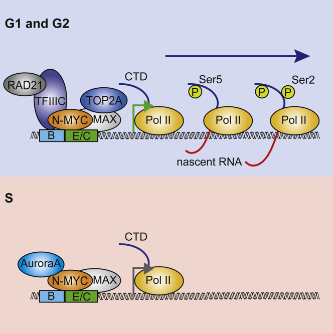
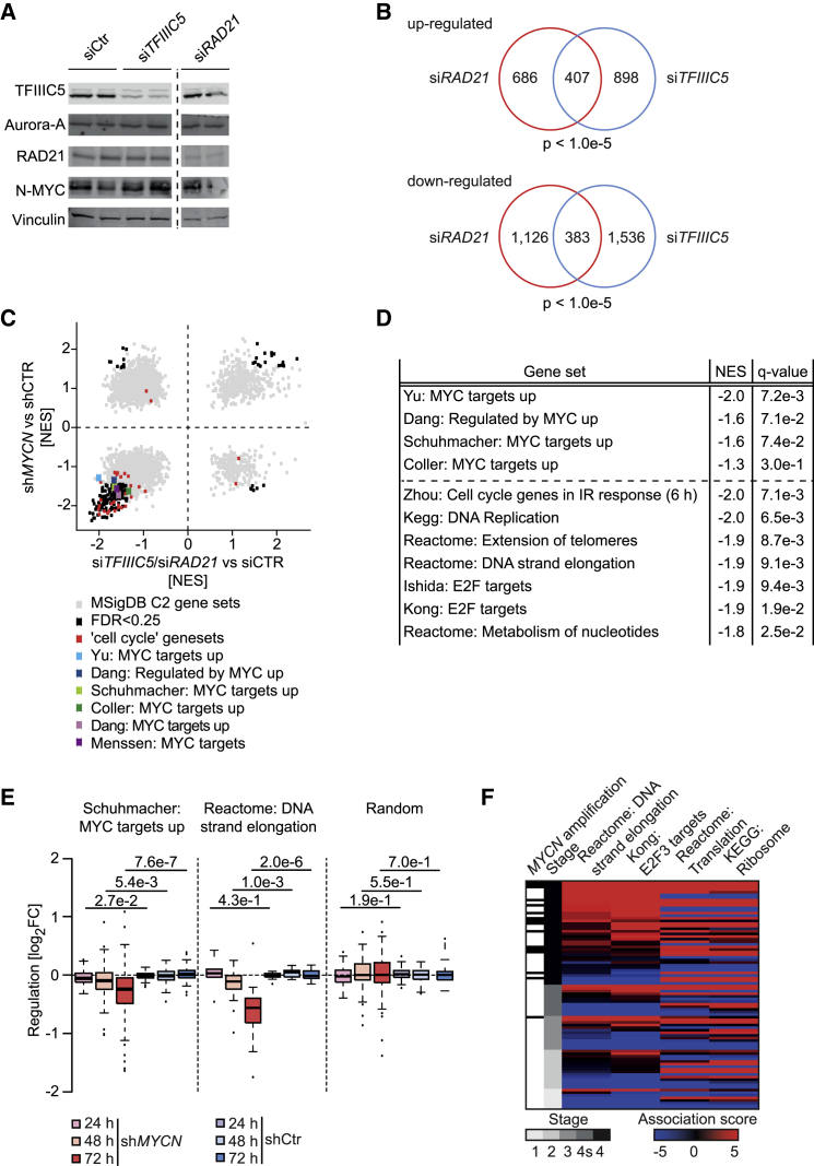
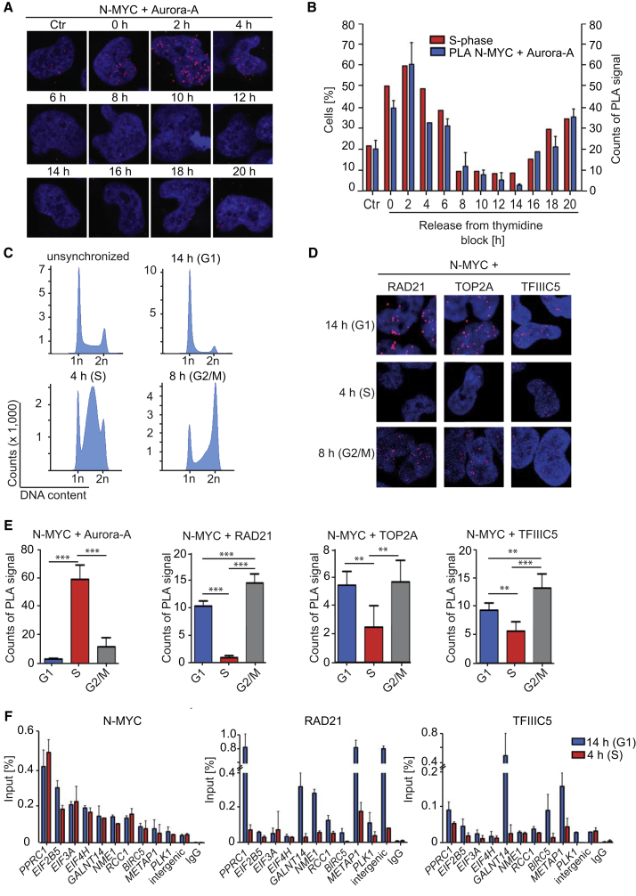
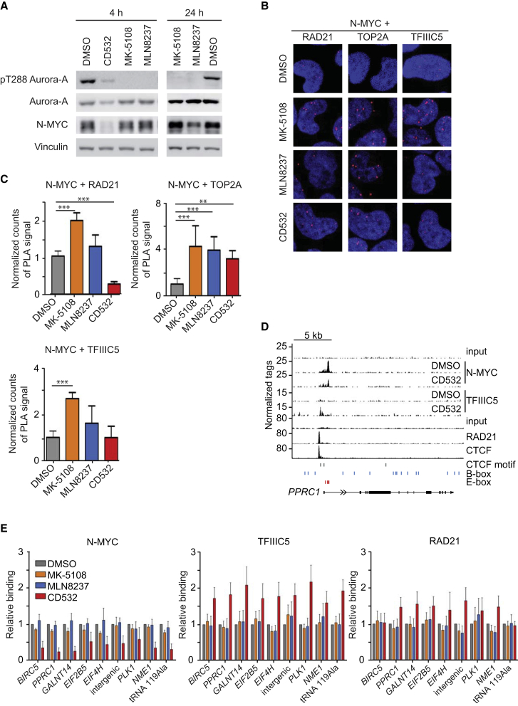
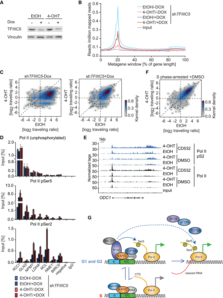
MYC proteins bind globally to active promoters and promote transcriptional elongation by RNA polymerase II (Pol II). To identify effector proteins that mediate this function, we performed mass spectrometry on N-MYC complexes in neuroblastoma cells. The analysis shows that N-MYC forms complexes with TFIIIC, TOP2A, and RAD21, a subunit of cohesin. N-MYC and TFIIIC bind to overlapping sites in thousands of Pol II promoters and intergenic regions. TFIIIC promotes association of RAD21 with N-MYC target sites and is required for N-MYC-dependent promoter escape and pause release of Pol II. Aurora-A competes with binding of TFIIIC and RAD21 to N-MYC in vitro and antagonizes association of TOP2A, TFIIIC, and RAD21 with N-MYC during S phase, blocking N-MYC-dependent release of Pol II from the promoter. Inhibition of Aurora-A in S phase restores RAD21 and TFIIIC binding to chromatin and partially restores N-MYC-dependent transcriptional elongation. We propose that complex formation with Aurora-A controls N-MYC function during the cell cycle.

Keywords: Aurora-A; MYC; N-MYC; RAD21; TFIIIC; neuroblastoma; pause release.


Figures

None
Graphical abstract
Figure 1
Figure 1
Complexes of N-MYC with TFIIIC, TOP2A, RAD21, and Aurora-A (A) Immunoblot documenting levels of ectopically expressed (exo) N-MYCwt and N-MYCmut proteins in stably infected SH-EP neuroblastoma cells relative to endogenous N-MYC (endo) of IMR-32 MYCN-amplified neuroblastoma cells. Where indicated, ectopically expressed proteins carry an N-terminal HA tag (NT HA); hence, their molecular weight is slightly larger than that of the endogenous protein (n = 2). (B) Results of mass spectrometry of α-HA immunoprecipitates of N-MYCwt and N-MYCmut complexes. The axes show the normalized ratio of peptides recovered in an α-HA immunoprecipitation from cells expressing N-MYCwt or N-MYCmut relative to an α-HA immunoprecipitation (IP) from control cells. Dot size represents the MaxQuant protein scores, which indicates the reliability of protein identification (Cox and Mann, 2008). (C) Immunoblots of α-N-MYC (left) and α-TFIIIC5 (right) immunoprecipitates from IMR-32 cells. The input corresponds to 1% of the amount used for the precipitation. Where indicated, ethidium bromide (EtBr) was added to a final concentration of 1 μg ml−1 to disrupt DNA-dependent interactions. Non-specific immunoglobulin G (IgG) was used for control immunoprecipitations (n = 3). (D) Pull-down assays from cell lysates documenting binding of TFIIIC5 and RAD21 to FLAG-tagged peptides spanning the indicated amino acids of the N-MYC N terminus. The input corresponds to 0.6% of the amount used for the precipitation (n = 2). The graph at the bottom visualizes the binding of the different N-MYC peptides. I/II indicate N-MYC sequences that mediate binding. (E) Immunoblots of α-N-MYC (left) and α-Aurora-A (right) immunoprecipitates from MYCN-amplified IMR-5 cells. The input corresponds to 1% of the amount used for the precipitation. Non-specific IgG was used for control immunoprecipitates (n = 4). (F) Pull-down assays from cell lysates documenting binding of TFIIIC5 and RAD21 to FLAG-tagged N-MYC peptides spanning amino acids 1–137 upon competition with Aurora-A. Recombinant Aurora-A protein was added in a concentration-dependent manner from 0.25 to 5 molar equivalents (Aurora-A/N-MYC peptide). 5 molar equivalents of glutathione-S-transferase (GST) were used as a control (n = 3). See also Figure S1 and Table S1. n indicates the number of independent biological replicas for each experiment.
Figure 2
Figure 2
Chromatin Binding of N-MYC/TFIIIC Complexes (A) Genome browser tracks at the NME1 locus illustrating chromatin association of the indicated proteins. The positions of B- and E-boxes and of CTCF motifs are indicated by vertical lines. The upper input is for ChIP sequencing of N-MYC and TFIIIC5; the lower input is for RAD21 and CTCF. (B) Top: Venn diagram documenting genome-wide overlap of N-MYC and TFIIIC5 binding sites in IMR-5 neuroblastoma cells. The p value was calculated using a permutation test. Bottom: diagram showing the location of N-MYC/TFIIIC5 sites in the genome. (C) De novo motif search in N-MYC- and/or TFIIIC5-bound regions. In overlapping sites, both peak regions were analyzed. The numbers indicate the percentage of sites in which the indicated motif was found. E values for enrichment of the respective motif are shown in Figure S2D. Motifs are only shown if the enrichment was significant. (D) Central enrichment of E-box, CTCF, and AP2a (as a negative control) motifs in the N-MYC peak of N-MYC/TFIIIC5 joint sites in Pol II promoters. The E value is calculated by a binominal test and adjusted for the number of motifs tested. (E) Heatmap showing occupancy of N-MYC, TFIIIC5, RAD21, and CTCF on overlapping N-MYC/TFIIIC sites in IMR-5 cells. Samples are normalized to the same number of mapped reads, and peaks are sorted according to N-MYC binding. (F) Boxplot documenting occupancy of the indicated proteins at joint N-MYC/TFIIIC5 binding sites (n = 1,630) and at N-MYC binding sites lacking TFIIIC5 (n = 2,406) located in promoters of Pol II genes. The number of reads was counted in a region of ± 100 bp around the N-MYC peak summit. See also Figure S2. n indicates the number of independent biological replicas for each experiment.
Figure 3
Figure 3
Assembly of N-MYC/TFIIIC and RAD21 Complexes on Chromatin (A) Venn diagram documenting genome-wide overlap of N-MYC/TFIIIC5 joint binding sites with RAD21 binding sites. The p value was calculated using a permutation test with 100,000 iterations. (B) Immunoblots of α-TFIIIC5 immunoprecipitates from IMR-5 cells. The input corresponds to 1% of the amount used for the precipitation. Non-specific IgG was used for control immunoprecipitates. Where indicated, CD532 (1 μM) was added to cells 4 hr prior to immunoprecipitation (n = 3). (C) Immunoblot showing levels of the indicated proteins in response to depletion of TFIIIC5. IMR-5 cells expressing an inducible shRNA directed against TFIIIC5 were treated with doxycycline (Dox) for 48 hr or with ethanol (EtOH) as a control (n = 3). (D) ChIP experiments documenting binding of TFIIIC5, N-MYC, and RAD21 to the indicated loci upon depletion of TFIIIC5. Error bars show SD of technical triplicates from one experiment (n = 2). (E) Immunoblot showing levels of the indicated proteins in response to depletion of N-MYC. IMR-5 cells expressing an inducible shRNA directed against N-MYC were treated with Dox for 48 hr or with EtOH as a control (n = 3). (F) ChIP experiments documenting binding of TFIIIC5, N-MYC, and RAD21 to the indicated loci upon depletion of N-MYC. Error bars show SD of technical triplicates from one experiment (n = 2). See also Figure S3. n indicates the number of independent biological replicas for each experiment.
Figure 4
Figure 4
Gene Regulation by N-MYC, TFIIIC, and RAD21 (A) Immunoblots documenting levels of the indicated proteins 48 hr after transfection of specific siRNAs. Duplicate samples are shown, both of which were used for RNA sequencing. All lanes are from the same exposure of a single immunoblot. (B) Venn diagrams documenting the overlap of upregulated (top) and downregulated (bottom) genes after depletion of TFIIIC5 or RAD21 in IMR-5 neuroblastoma cells. The p values were calculated using a Monte Carlo simulation with 100,000 permutations and all expressed genes (n = 17,450) as the basis. (C) Correlation of gene sets that change in expression upon depletion of N-MYC with the aggregate of changes in response to siRNA-mediated depletion of RAD21 and TFIIIC5. Each dot reflects a gene set. A light gray color indicates that the expression change of a gene set was not statistically significant. Published sets of MYC target genes are colored. (D) Examples of gene sets that are downregulated in response to depletion of both RAD21 and TFIIIC5. NES is the normalized enrichment score, indicating direction and extent of regulation. (E) Boxplots documenting changes in expression of selected gene sets upon depletion of N-MYC using a Dox-inducible shRNA in IMR-32 neuroblastoma cells. (F) Heatmap illustrating stage-specific expression of N-MYC/TFIIIC/RAD21-regulated genes sets in human neuroblastoma cells. The black bars in the first row indicate MYCN amplification status. See also Figure S4. n indicates the number of independent biological replicas for each experiment.
Figure 5
Figure 5
Regulation of N-MYC Transcription Complexes during the Cell Cycle (A) Representative pictures from proximity ligation assays (PLAs) documenting complex formation between N-MYC and Aurora-A in IMR-5 cells after release from a double thymidine block. Non-synchronized cells are shown as a control (Ctr). Nuclei were stained using Hoechst. Red dots show PLA signals resulting from N-MYC/Aurora-A interactions (n = 3). (B) Quantification of the PLA shown in (A). The percentage of cells in S phase is indicated in parallel. Error bars show SD of technical triplicates from one representative experiment (n = 3). (C) Representative FACS profiles of propidium iodide (PI)-stained cells documenting cell cycle distribution at the indicated times after release from a double thymidine block. (D) Representative pictures from PLAs documenting complex formation between N-MYC and RAD21 and TOP2A and TFIIIC5 in IMR-5 cells at the indicated times after release from a double thymidine block. Nuclei were stained using Hoechst. Red dots show PLA signals (n = 3). (E) Quantification of PLAs shown in (A) and (D). Bars show mean + SD of technical triplicates from one representative experiment (n = 3). ∗∗p < 0.01, ∗∗∗p < 0.001. (F) ChIP of IMR-5 cells documenting chromatin association of N-MYC, RAD21, and TFIIIC5 at the indicated gene loci at the indicated times after release from a double thymidine block. Error bars show SD of technical triplicates from one representative experiment (n = 3). See also Figure S5. n indicates the number of independent biological replicas for each experiment.
Figure 6
Figure 6
Role of Aurora-A in Dynamics of N-MYC Complexes during the Cell Cycle (A) Immunoblot documenting levels of the indicated proteins and of Aurora-A, which is autophosphorylated at T288 (indicating catalytically active Aurora-A), in IMR-5 MYCN-amplified neuroblastoma cells after 4 hr (left) or 24 hr (right) exposure to 1 μM of the indicated Aurora-A inhibitors (n = 4). (B) Representative pictures from PLAs documenting complex formation between N-MYC and RAD21 and TOP2A or TFIIIC5 in IMR-5 cells released for 4 hr from a double thymidine block in the presence of the indicated Aurora-A inhibitors (1 μM) or DMSO as a control. Nuclei were stained using Hoechst. Red dots show signals arising from interaction of N-MYC with the indicated proteins. (C) Quantification of PLAs shown in (B). Data are normalized to DMSO-treated cells. Bars show mean + SD of technical triplicates from one experiment (n = 4). ∗∗p < 0.01, ∗∗∗p < 0.001. (D) Genome browser tracks at the PPRC1 locus, illustrating chromatin association of the indicated proteins. The positions of B- and E-boxes and of CTCF motifs are indicated by vertical lines. The upper input is for ChIP sequencing of N-MYC and TFIIIC5, the lower input is for ChIP-sequencing of RAD21 and CTCF. (E) ChIP of IMR-5 cells documenting chromatin association of N-MYC, TFIIIC5, and RAD21 at the indicated loci after treatment (1 μM) with MK-5108 (24 hr), MLN8237 (24 hr), CD532 (4 hr), or DMSO as a control. Error bars show SEM of three independent experiments. Data are normalized to DMSO-treated cells. See also Figure S6. n indicates the number of independent biological replicas for each experiment.
Figure 7
Figure 7
Aurora-A Suppresses N-MYC-Dependent Pause Release of Pol II in S phase (A) Immunoblot showing levels of TFIIIC5 in SH-EP-N-MYCER cells expressing a Dox-inducible shRNA targeting TFIIIC5 (n = 3). Dox (1 μg ml−1) was added for 30 hr; EtOH was used as a control. (B) Metagene plot of all expressed genes (n = 14,650) illustrating distribution of Pol II within transcribed regions before and 5 hr after activation of N-MYCER in cells expressing Dox-inducible shTFIIIC5. (C) 2D kernel density plot showing the ratio of Pol II occupancy at the promoter to occupancy in the gene body (traveling ratio) in cells treated as above for all expressed genes (n = 14,650) before and after 5 hr of N-MYCER-activation. (D). ChIP of SH-EP N-MYCER cells documenting chromatin association of hypo-phosphorylated Pol II and Pol II phosphorylated at Ser5 (pSer5) or Ser2 (pSer2) at the indicated gene loci before and 5 hr after activation of N-MYCER. Occupancy at the transcription start site (TSS) was analyzed for hypo-phosphorylated Pol II and pSer5Pol II. Occupancy at the transcription end site (TES) was analyzed for pSer2Pol II. Dox (1 μg ml−1) was added for 30 hr; EtOH was used as a control. Error bars show SD of technical triplicates from one representative experiment (n = 3). (E) Genome browser tracks illustrating chromatin association of Pol II and Pol II pSer2 at the ODC1 locus. ChIP sequencing was performed on cells synchronized in S phase by double thymidine blockade and treated for 2 hr with CD532 where indicated (1 μM). (F) 2D kernel density plot showing the Pol II traveling ratio in response to N-MYCER activation in SH-EP cells that were synchronized in S phase by double thymidine block for all expressed genes (n = 14,650) before and after 4 hr of N-MYCER activation. (G) Model summarizing our findings. We propose that the multiple protein-protein interactions of N-MYC promote sequential phosphorylation events of Pol II and both promotor escape and pause release. The dashed line indicates that effects on pause release and Ser2 phosphorylation could be secondary to changes in Ser5 phosphorylation. E/C indicates that the complex associates with sites on chromatin that contain either an E-box or a CTCF motif. See also Figure S7. n indicates the number of independent biological replicas for each experiment.

References

    1. Baranello L., Wojtowicz D., Cui K., Devaiah B.N., Chung H.J., Chan-Salis K.Y., Guha R., Wilson K., Zhang X., Zhang H. RNA polymerase II regulates topoisomerase 1 activity to favor efficient transcription. Cell. 2016;165:357–371. - PMC - PubMed
    1. Beltran H. The N-MYC oncogene: maximizing its targets, regulation, and therapeutic potential. Mol. Cancer Res. 2014;12:815–822. - PubMed
    1. Blackwell T.K., Huang J., Ma A., Kretzner L., Alt F.W., Eisenman R.N., Weintraub H. Binding of MYC proteins to canonical and noncanonical DNA sequences. Mol. Cell. Biol. 1993;13:5216–5224. - PMC - PubMed
    1. Carrière L., Graziani S., Alibert O., Ghavi-Helm Y., Boussouar F., Humbertclaude H., Jounier S., Aude J.C., Keime C., Murvai J. Genomic binding of Pol III transcription machinery and relationship with TFIIS transcription factor distribution in mouse embryonic stem cells. Nucleic Acids Res. 2012;40:270–283. - PMC - PubMed
    1. Chesler L., Schlieve C., Goldenberg D.D., Kenney A., Kim G., McMillan A., Matthay K.K., Rowitch D., Weiss W.A. Inhibition of phosphatidylinositol 3-kinase destabilizes MYCN protein and blocks malignant progression in neuroblastoma. Cancer Res. 2006;66:8139–8146. - PMC - PubMed