Review
. 2017 Dec 19;113(12):2579-2594.
doi: 10.1016/j.bpj.2017.09.037.

Regulation of Contraction by the Thick Filaments in Skeletal Muscle

Affiliations
Review

Regulation of Contraction by the Thick Filaments in Skeletal Muscle

Malcolm Irving. Biophys J. .

Abstract

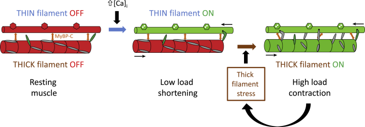
Contraction of skeletal muscle cells is initiated by a well-known signaling pathway. An action potential in a motor nerve triggers an action potential in a muscle cell membrane, a transient increase of intracellular calcium concentration, binding of calcium to troponin in the actin-containing thin filaments, and a structural change in the thin filaments that allows myosin motors from the thick filaments to bind to actin and generate force. This calcium/thin filament mediated pathway provides the "START" signal for contraction, but it is argued that the functional response of the muscle cell, including the speed of its contraction and relaxation, adaptation to the external load, and the metabolic cost of contraction is largely determined by additional mechanisms. This review considers the role of the thick filaments in those mechanisms, and puts forward a paradigm for the control of contraction in skeletal muscle in which both the thick and thin filaments have a regulatory function. The OFF state of the thick filament is characterized by helical packing of most of the myosin head or motor domains on the thick filament surface in a conformation that makes them unavailable for actin binding or ATP hydrolysis, although a small fraction of the myosin heads are constitutively ON. The availability of the majority fraction of the myosin heads for contraction is controlled in part by the external load on the muscle, so that these heads only attach to actin and hydrolyze ATP when they are required. This phenomenon seems to be the major determinant of the well-known force-velocity relationship of muscle, and controls the metabolic cost of contraction. The regulatory state of the thick filament also seems to control the dynamics of both muscle activation and relaxation.


Figures

Figure 1
Figure 1
Arrangement of the thick (red, orange) and thin (blue) filaments in the muscle sarcomere. The midpoints of the thick filaments are connected by the M-line (dark red) and the ends of the thin filaments by the Z-line (black), which is considered to define the ends of the sarcomere. The central third of each half thick filament is the myosin binding protein C-containing C zone (orange). Titin (brown) runs from the M-line to the Z-line. The white arrows indicate the forces imposed on the Z and M lines during contraction, and the reversal of filament polarity at those lines. To see this figure in color, go online.
Figure 2
Figure 2
(A) Schematic model of possible structures of the thin and thick filaments in the sarcomeric C zone in the OFF state. The thin filaments (drawn from coordinates in PDB: 2W4U) have a helical periodicity of ∼38 nm and contain actin (light and dark gray), tropomyosin (purple) and troponin (yellow). The thick filaments have a helical periodicity of ∼43 nm, with three myosin molecules, each with a pair of myosin heads in the interacting heads motif (IHM), every 14.3 nm. The single IHM outlined in the gray ellipse is shown in greater detail in (B). Titin (brown; based on PDB: 3LCY) and the C terminus of myosin binding protein-C (orange; based on PDB: 5K6P and 2EDN) are shown in the approximate conformations suggested by electron microscopy of isolated filaments (46, 47); the N terminus of MyBP-C is shown bound to actin. The detailed structural relationships between the thick filament components, and the organization of the long C-terminal regions of the myosin tails in the filament backbone are unknown; the latter is shown as a simple cylinder. (B). The IHM thought to correspond to the OFF conformation of the myosin heads on the surface of the thick filament. Myosin heavy chain (MHC), red; essential light chain (ELC), green; regulatory light chain (RLC), blue; with lighter and darker shades corresponding to the two myosin heads in each myosin molecule (free and blocked heads. respectively), folding back onto the coiled-coil S) tail. Coordinates from PDB: 3DTP. To see this figure in color, go online.
Figure 3
Figure 3
(A) Dependence on steady [Ca2+], plotted as pCa= −log10 [Ca2+], of isometric force (solid triangles) and the orientation of a fluorescent probe attached to the RLC region of the myosin heads (〈P2〉, open triangles), replotted from (68). (B) Time course of [Ca2+] and isometric force after single action potential stimulation, from (82). (C) Time course of the intensity of the first myosin-based x-ray layer line (IML1; solid circles) signaling the helical order of the myosin heads on the surface of the thick filaments and force (solid line) during the rising phase of an isometric tetanus, replotted from (61). Time zero and the vertical line mark the first electrical stimulus. (D). Time course of IML1 (circles) and force (solid line) during isometric relaxation (solid circles) and chaotic relaxation (open circles), replotted from (111). The full vertical line and time zero mark the last electrical stimulus; the dashed vertical line the transition between isometric and chaotic relaxation; the dashed horizontal line is IML1 in resting muscle. Data in (A) from skinned fibers of rabbit psoas muscle, 25°C; those in (BD) from intact fibers of frog fast twitch muscle, 4°C.
Figure 4
Figure 4
Dual filament regulation in skeletal muscle. The thin filament (upper) is shown as a simple cylinder with hexagons symbolizing troponin; the thin filament is switched ON by calcium binding (blue arrow). Part of the C zone of the thick filament (lower) is shown with a surface array of myosin head domains (gray or green); the thick filament can be switched ON by mechanical stress (brown arrow). OFF and ON states of both filaments are red and green respectively. Constitutively ON myosin heads are green; myosin heads under thick filament control are gray. The double-headed orange arrows symbolize coupling of the regulatory states of the thin and thick filaments by myosin binding protein-C, which may have a different conformation in the OFF and ON states of the thick filament. To see this figure in color, go online.

References

    1. Woledge R.C., Curtin N.A., Homsher E. Academic Press; London, UK: 1985. Energetic Aspects of Muscle Contraction. - PubMed
    1. Cooke R. The role of the myosin ATPase activity in adaptive thermogenesis by skeletal muscle. Biophys. Rev. 2011;3:33–45. - PMC - PubMed
    1. Squire J.M. Plenum Press; New York: 1981. The Structural Basis of Muscle Contraction.
    1. Huxley H.E. The mechanism of muscular contraction. Science. 1969;164:1356–1365. - PubMed
    1. Huxley A.F. Muscular contraction. J. Physiol. 1974;243:1–43. - PMC - PubMed