. 2017 Oct 6;8(61):103581-103600.
doi: 10.18632/oncotarget.21658. eCollection 2017 Nov 28.

Human papillomavirus type 18 E5 oncogene supports cell cycle progression and impairs epithelial differentiation by modulating growth factor receptor signalling during the virus life cycle

Affiliations

Human papillomavirus type 18 E5 oncogene supports cell cycle progression and impairs epithelial differentiation by modulating growth factor receptor signalling during the virus life cycle

Christopher W Wasson et al. Oncotarget. .

Abstract

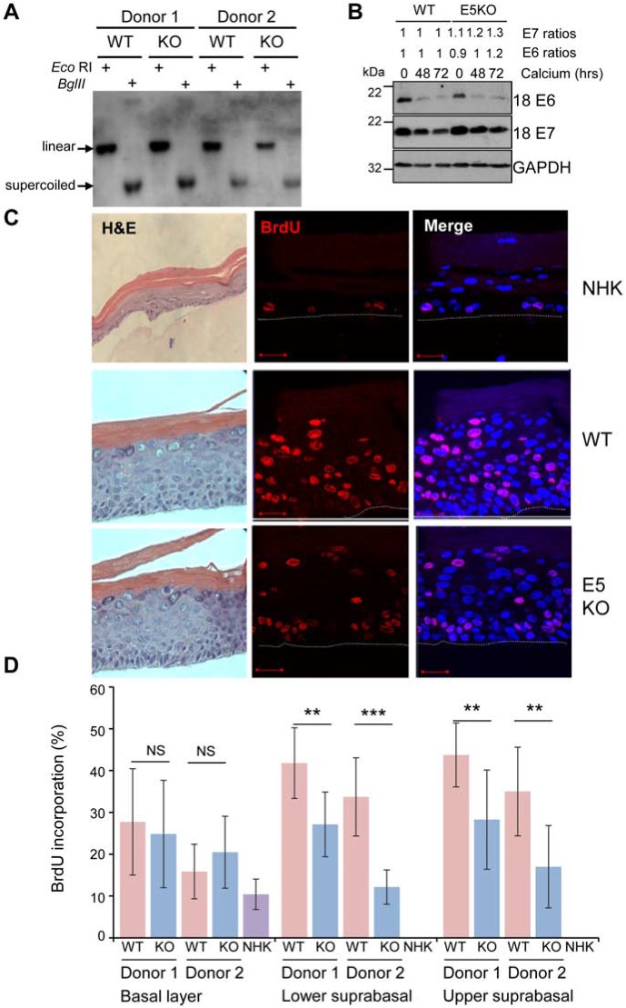
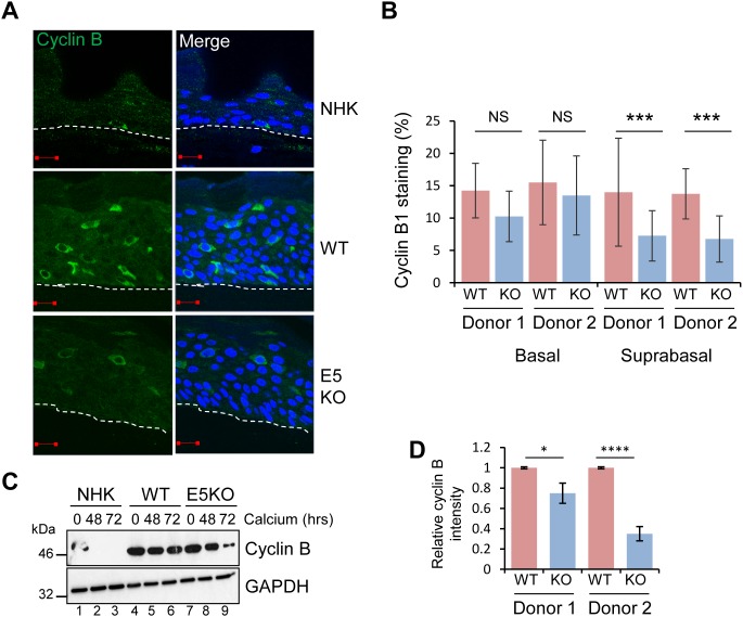
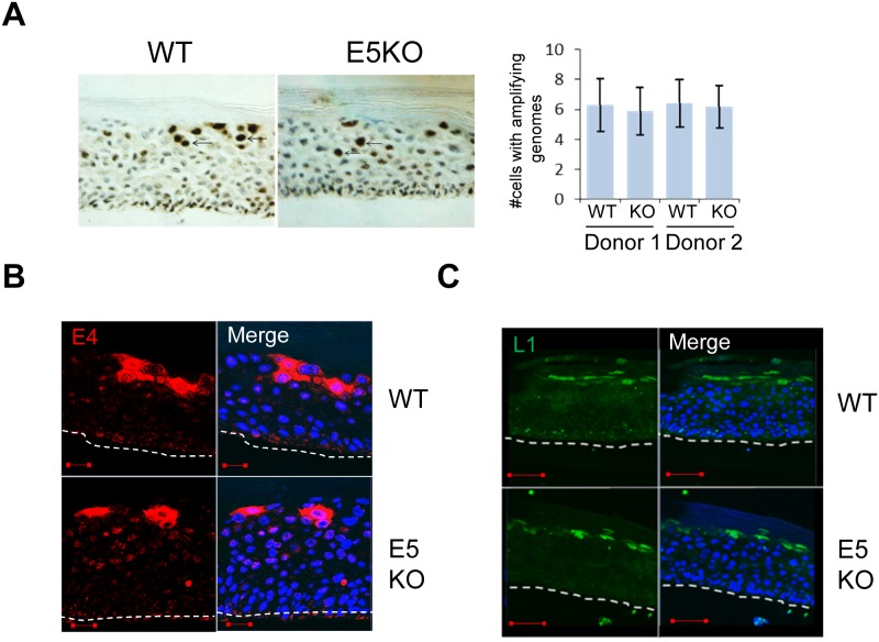
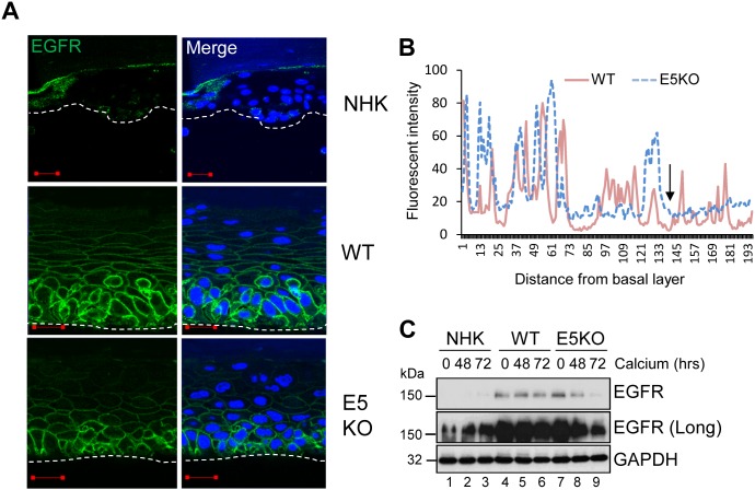
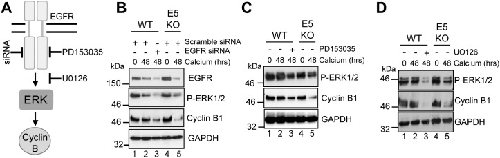
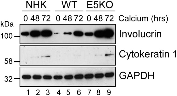
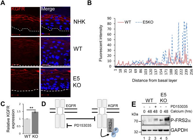
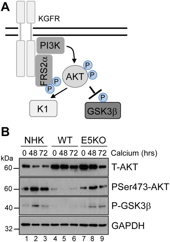
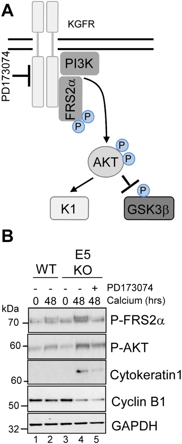
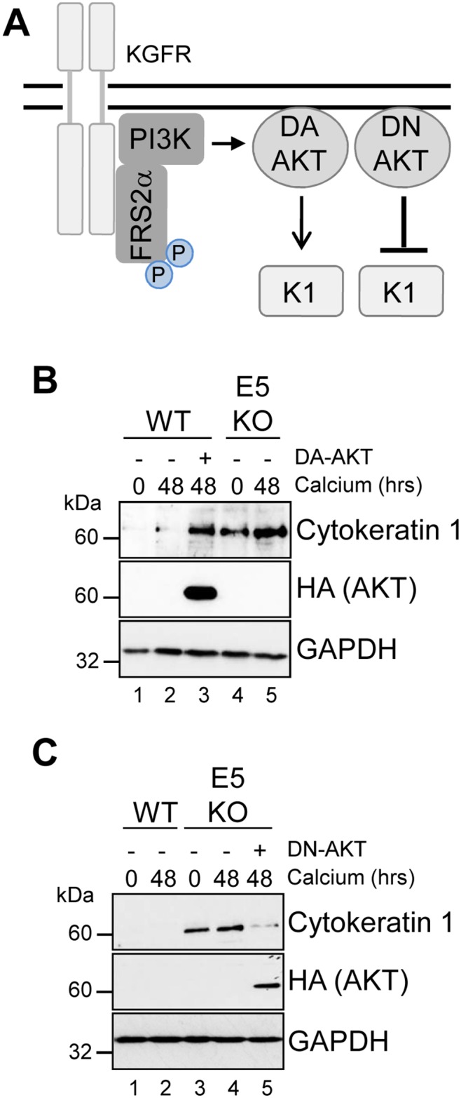
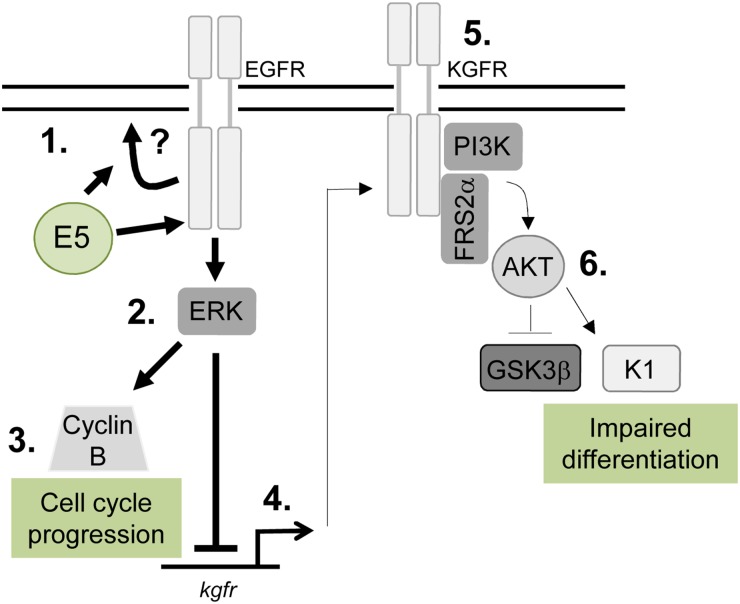
Deregulation of proliferation and differentiation-dependent signalling pathways is a hallmark of human papillomavirus (HPV) infection. Although the manipulation of these pathways by E6 and E7 has been extensively studied, controversies surround the role of the E5 oncoprotein during a productive virus life cycle. By integrating primary keratinocytes harbouring wild type or E5 knockout HPV18 genomes with pharmacological and gain/loss of function models, this study aimed to provide molecular information about the role of E5 in epithelial proliferation and differentiation. We show that E5 contributes to cell cycle progression and unscheduled host DNA synthesis in differentiating keratinocytes. E5 function correlates with increased EGFR activation in differentiating cells and blockade of this pathway impairs differentiation-dependent cell cycle progression of HPV18 containing cells. Our findings provide a functional requirement of enhanced EGFR signalling for suprabasal cellular DNA synthesis during the virus life cycle. They also reveal an unrecognised contribution of E5 towards the impaired keratinocyte differentiation observed during a productive HPV infection. E5 suppresses a signalling axis consisting of the keratinocyte growth factor receptor (KGFR) pathway. Inhibition of this pathway compensates for the loss of E5 in knockout cells and re-instates the delay in differentiation. The negative regulation of KGFR involves suppression by the EGFR pathway. Thus our data reveal an unappreciated role for E5-mediated EGFR signalling in orchestrating the balance between proliferation and differentiation in suprabasal cells.

Keywords: E5; EGFR; HPV; differentiation; proliferation.


Conflict of interest statement

CONFLICTS OF INTEREST The authors declare that there is no conflict of interest.

Figures

Figure 1
Figure 1. HPV18 E5 contributes towards unscheduled host cell DNA synthesis upon keratinocyte differentiation
(A) Southern analysis of equal amounts of total DNA extracted from NHK transfected with wild-type (WT) or mutant (E5KO) genomes in two different donors. DNA was digested with DpnI (digests input DNA) together with BglII which does not digest HPV18 DNA or EcoRI which linearizes HPV18 genomes. (B) Detection of E6 and E7 proteins in equal amounts of Triton-X100 detergent soluble protein lysates prepared from cells differentiated in high-calcium monolayer cultures. Levels of GAPDH were used as a loading control. Densitometry analysis of protein bands was performed using ImageJ software. (C) Organotypic rafts were incubated with BrdU to identify nuclei positive for cellular DNA synthesis. BrdU positive cells were visualised with an anti-BrdU antibody (red) and nuclei visualised with DAPI (blue). White dotted lines indicate the basal cell layer. Red scale bar represents 20 μm. Organotypic raft cultures were also stained with haematoxylin and eosin to observe the gross morphology of the epithelium. (D) Graphs showing the percentage of BrdU positive nuclei in basal and lower suprabasal and upper suprabasal layers. The data, shown as a mean with standard deviation, were derived from 15 fields of view of each raft and from 3 independent experiments from two donors cell lines. Significance as determined by Student’s t-test is shown as ** = p<0.01, *** = p<0.001.
Figure 2
Figure 2. HPV18 E5 deregulates cell cycle progression in differentiated cells
(A) Organotypic rafts stained for cyclin B1 (green) and nuclei stained with DAPI (blue), white dotted lines indicate the basal cell layer. Red scale bar represents 20 μm. (B) Graphs showing the percentage of cyclin B1 positive nuclei in basal and suprabasal layers. The data, shown as a mean with standard deviation, were derived from 15 fields of view of each raft and from 3 independent experiments from two donor cell lines. Significance as determined by Student’s t-test is shown as *** = p<0.001. (C) Lysates of NHK, WT and E5KO cells subjected to high calcium differentiation were analysed for cyclin B1 expression by immunoblotting. GAPDH served as a loading control. (D) Graph showing densitometry analysis of cyclin B1 expression at 72 hours for two donors. The data is shown as a mean and standard deviation from three independent experiments. Significance as determined by Student’s t-test is shown as * = p<0.05 and **** = p<0.0001.
Figure 3
Figure 3. HPV18 E5 function is not necessary for genome amplification and late protein expression in differentiating keratinocytes
(A) Organotypic raft cultures were probed with a biotin-conjugated HPV DNA specific for the high-risk HPV types to visualise genome amplification. Arrows indicate examples of CISH positive nuclei. Graph represents the mean (± standard deviation) of CISH positive nuclei per field of view (15 fields of view of each raft) for two separate donors. (B and C) Organotypic rafts stained for E4 (red) and L1 (green), nuclei were visualised with DAPI (blue). White dotted lines indicate the basal cell layer. Red scale bar represents 20 μm. Images are representative of staining from two donors and three independent experiments.
Figure 4
Figure 4. HPV18 E5 maintains EGFR expression in differentiating keratinocytes
(A) Organotypic rafts stained for EGFR (green), with nuclei stained with DAPI (blue). White dotted lines indicate the basal cell layer. Red scale bar represents 20 μm. Images are representative of staining from two donors. (B) Histogram analysis of the staining intensity from WT and E5KO rafts. Data was derived from 5 fields of view from three independent experiments and two donors. (C) Lysates of keratinocytes differentiated in high calcium media analysed for total EGFR expression by immunoblotting. GAPDH served as a loading control. Western blots are representative of three independent experiments in two donor lines.
Figure 5
Figure 5. HPV18 requires EGFR activation to maintain cyclin B1 expression in differentiating cells
(A) Schematic showing the EGFR/ERK signalling pathway and the targets of the siRNA and inhibitors used in this study. Mock treatedkeratinocytes were differentiated in high calcium media and lysed after 48 hours. In parallel, keratinocytes were treated with (B) scramble or EGFR specific siRNA, (C) an EGFR kinase inhibitor (2 μM PD153035), or (D) a Mek1/2 kinase inhibitor (20 μM UO126) during differentiation. All samples were analysed for cyclin B1 and phosphorylated ERK1/2 expression. GAPDH served as a loading control. Representative blots are shown from at least three independent biological repeats from two donor lines.
Figure 6
Figure 6. HPV18 E5 impairs keratinocyte differentiation
Lysates of NHK, WT and E5KO keratinocytes subjected to high calcium differentiation were analysed for involucrin and cytokeratin 1 (K1) expression. GAPDH served as a loading control. Western blots are representative of three independent experiments in two donor lines.
Figure 7
Figure 7. HPV18 E5 inhibits the KGFR signalling pathway
(A) Organotypic raft cultures stained for KGFR expression (red) and nuclei stained with DAPI (blue). White dotted lines indicate the basal cell layer. Red scale bar represents 20 μm. Images are representative of staining from two donors. (B) Histogram analysis of the staining intensity from WT and E5KO rafts. Data was derived from 5 fields of view from three independent experiments, from two donors. (C) Graph showing relative KGFR mRNA expression in differentiated WT and E5KO keratinocytes after 96 hours incubation in high calcium media. Results were corrected for expression of an U6 loading control. The data is shown as a mean and standard deviation from three independent biological repeats from two donors. Significance as determined by Student’s t-test is shown as ** = p<0.01. (D) Schematic showing the interplay between the EGFR/KGFR signalling pathway and the target of the inhibitor used in this study. (E) Mock-treated keratinocytes were differentiated in high calcium media and lysed after 48 hours. In parallel, keratinocytes were treated with an EGFR kinase inhibitor (2 μM PD153035), during differentiation. All samples were analysed for phosphorylated FRS2α expression. GAPDH served as a loading control. Representative blots are shown from at least three independent biological repeats from two donor lines.
Figure 8
Figure 8. HPV18 E5 inhibits downstream targets of the KGFR signalling pathway
(A) Schematic showing the KGFR downstream signalling pathway including the KGFR substrate FRS2a and downstream kinases AKT and GSK3β. (B) Lysates of NHK, WT and E5KO keratinocytes subjected to high calcium differentiation were analysed for p-AKT, total AKT and p-GSK3β. GAPDH served as a loading control. Western blots are representative of three independent experiments in two donor lines
Figure 9
Figure 9. Pharmacological inhibition of KGFR activity compensates for loss of E5 and impairs keratinocyte differentiation
(A) Schematic showing the KGFR signalling pathway and target of the pharmacological inhibitor. (B) Control (vehicle only) keratinocytes were differentiated in high calcium media and lysed after 48 hours. Parallel cultures of E5KO keratinocytes were treated with 30 nM of a KGFR kinase inhibitor (PD173074). Lysates were analysed for the phosphorylated forms of FRS2α, AKT, GSK3α/β, cytokeratin 1 (K1) and cyclin B1 by immunoblot.
Figure 10
Figure 10. AKT is essential for the modulation of keratinocyte differentiation by HPV18
(A) Schematic showing the KGFR signalling pathway and the effects of the dominant active (DA) and negative (DN) AKT. HPV18 genome containing keratinocytes were infected with empty retrovirus, or with retroviruses encoding DA AKT (B) or DN AKT (C). Cells were differentiated in high calcium media for 48 hours prior to lysis and analysed for expression of cytokeratin 1 (K1) to assess differentiation. Expression of the exogenous AKT was confirmed using an antibody against the HA epitope and GAPDH served as a loading control.
Figure 11
Figure 11. Schematic model depicting the proposed role of EGFR signalling in the E5-mediated manipulation of proliferation and differentiation pathways during the virus life cycle
E5 expression results in enhanced EGFR surface expression and activation, through a process that might require endosome recycling (1). This increases ERK/MAPK activity (2), resulting in activation of substrates, which include cell cycle associated proteins e.g. cyclin B (3). EGFR can suppress keratinocyte differentiation, by inhibiting a number of targets including the KGFR pathway. KGFR transcription is suppressed in E5 expressing cells (4) and KGFR signalling is markedly reduced (5). As a consequence, targets of KGFR including Akt are suppressed, culminating in loss of expression of a number of spinous associated differentiation markers e.g. cytokeratin 1 (K1) (6). As a result keratinocyte differentiation is impaired in cells expressing E5.

References

    1. Doorbar J, Quint W, Banks L, Bravo IG, Stoler M, Broker TR, Stanley MA. The biology and life-cycle of human papillomaviruses. Vaccine. 2012;30:F55–F70. http://doi.org/10.1016/j.vaccine.2012.06.083. - DOI - PubMed
    1. Roberts S. Papillomaviruses. eLS. Chichester, UK: John Wiley & Sons, Ltd; 2001. pp. 1–11. http://doi.org/10.1002/9780470015902.a0000422.pub3. - DOI
    1. Clifford GM, Smith JS, Aguado T, Franceschi S. Comparison of HPV type distribution in high-grade cervical lesions and cervical cancer: a meta-analysis. Br J Cancer. 2003;89:101–105. http://doi.org/10.1038/sj.bjc.6601024. - DOI - PMC - PubMed
    1. de Sanjose S, Quint WG, Alemany L, Geraets DT, Klaustermeier JE, Lloveras B, Tous S, Felix A, Bravo LE, Shin HR, Vallejos CS, de Ruiz PA, Lima MA, et al. Human papillomavirus genotype attribution in invasive cervical cancer: a retrospective cross-sectional worldwide study. Lancet Oncol. 2010;11:1048–1056. http://doi.org/10.1016/S1470-2045(10)70230-8. - DOI - PubMed
    1. Dumesic PA, Scholl FA, Barragan DI, Khavari PA. Erk1/2 MAP kinases are required for epidermal G2/M progression. J Cell Biol. 2009;185:409–422. http://doi.org/10.1083/jcb.200804038. - DOI - PMC - PubMed