. 2017 Oct 23;8(61):103828-103842.
doi: 10.18632/oncotarget.21953. eCollection 2017 Nov 28.

LINE-1 couples EMT programming with acquisition of oncogenic phenotypes in human bronchial epithelial cells

Affiliations

LINE-1 couples EMT programming with acquisition of oncogenic phenotypes in human bronchial epithelial cells

Elsa M Reyes-Reyes et al. Oncotarget. .

Abstract

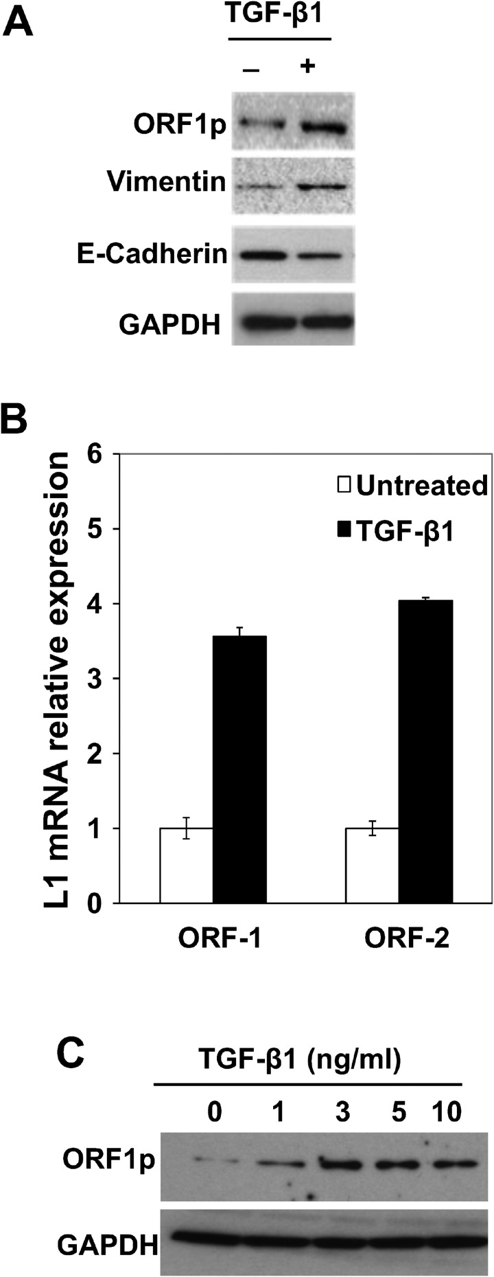
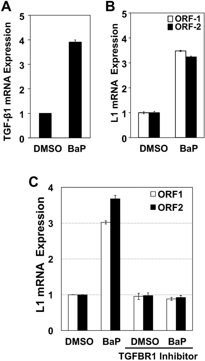
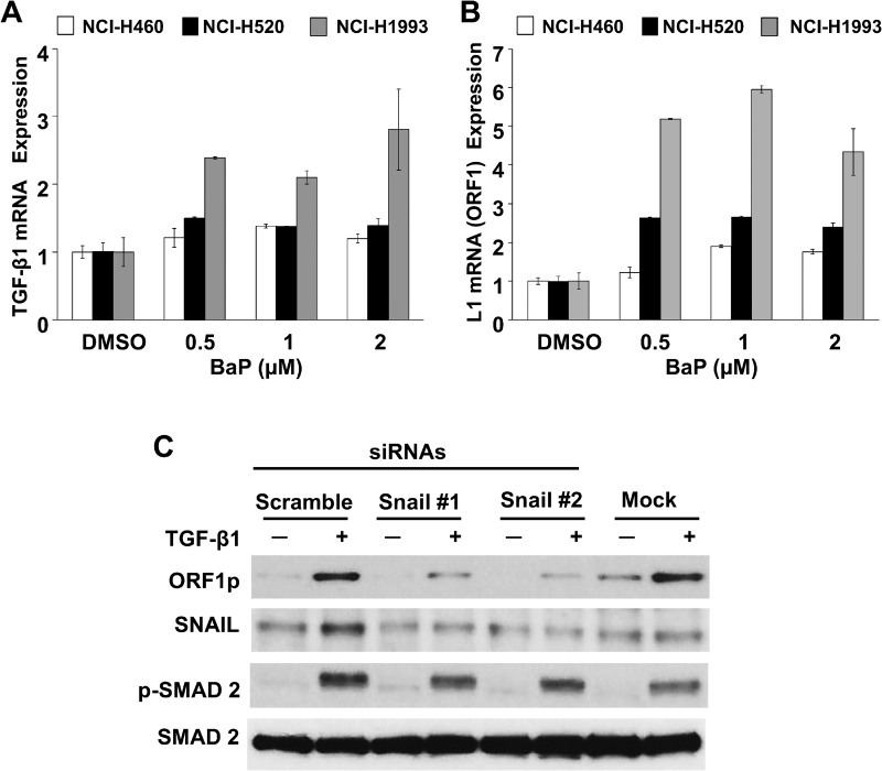
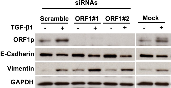
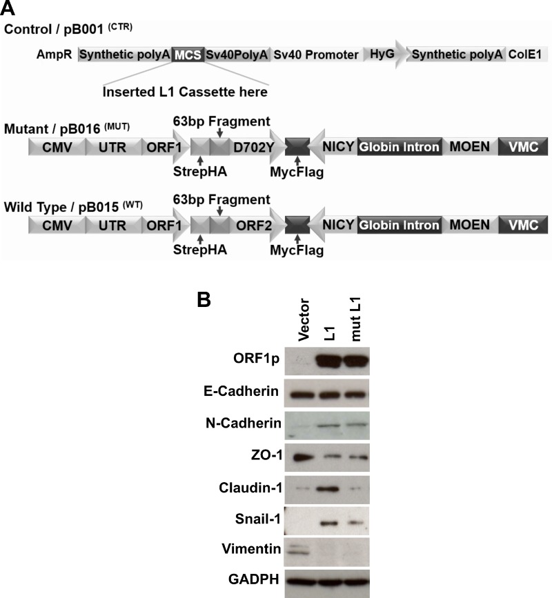
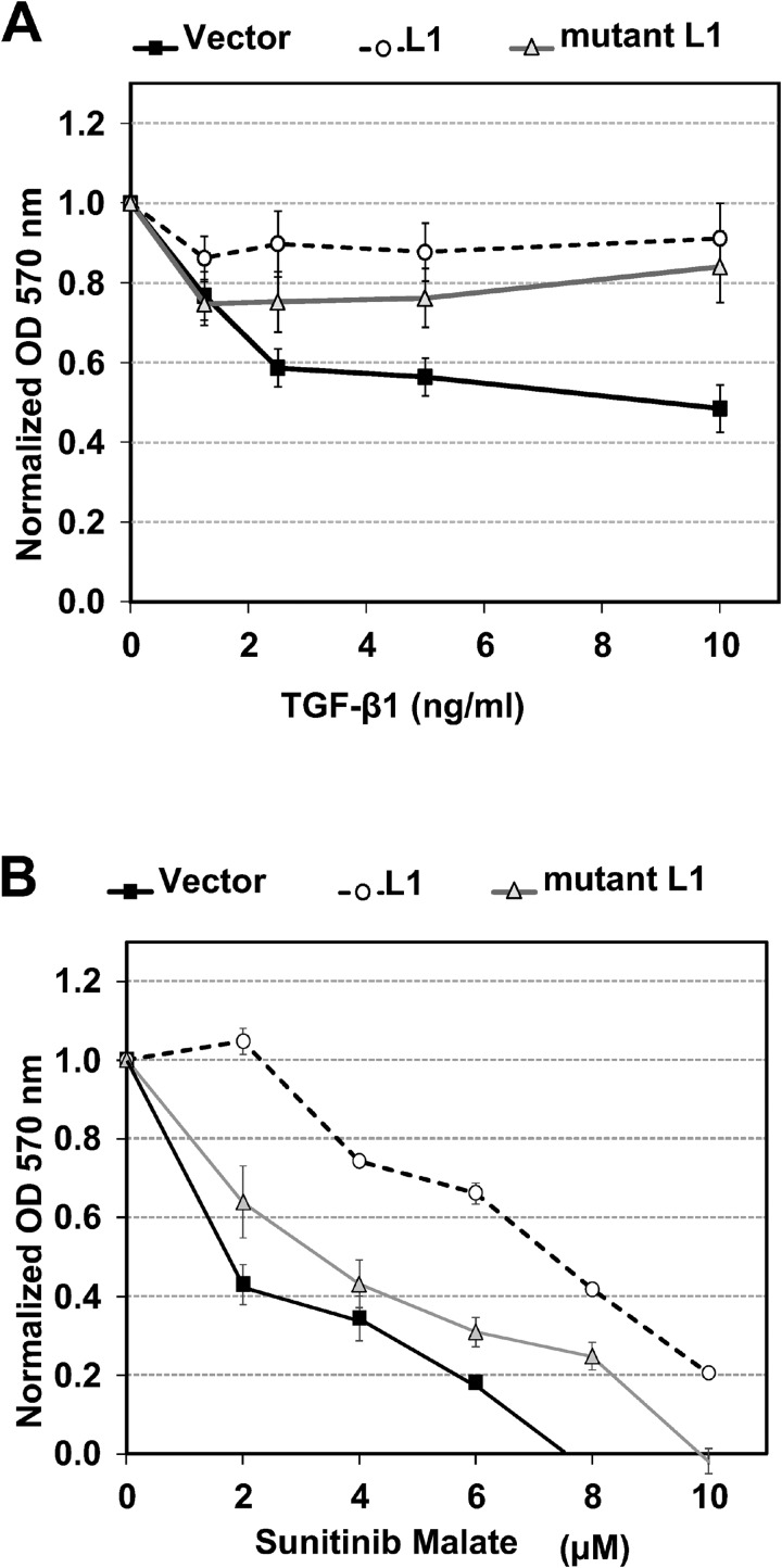
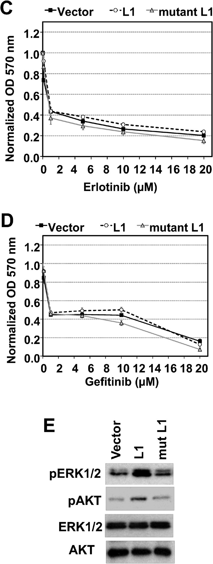
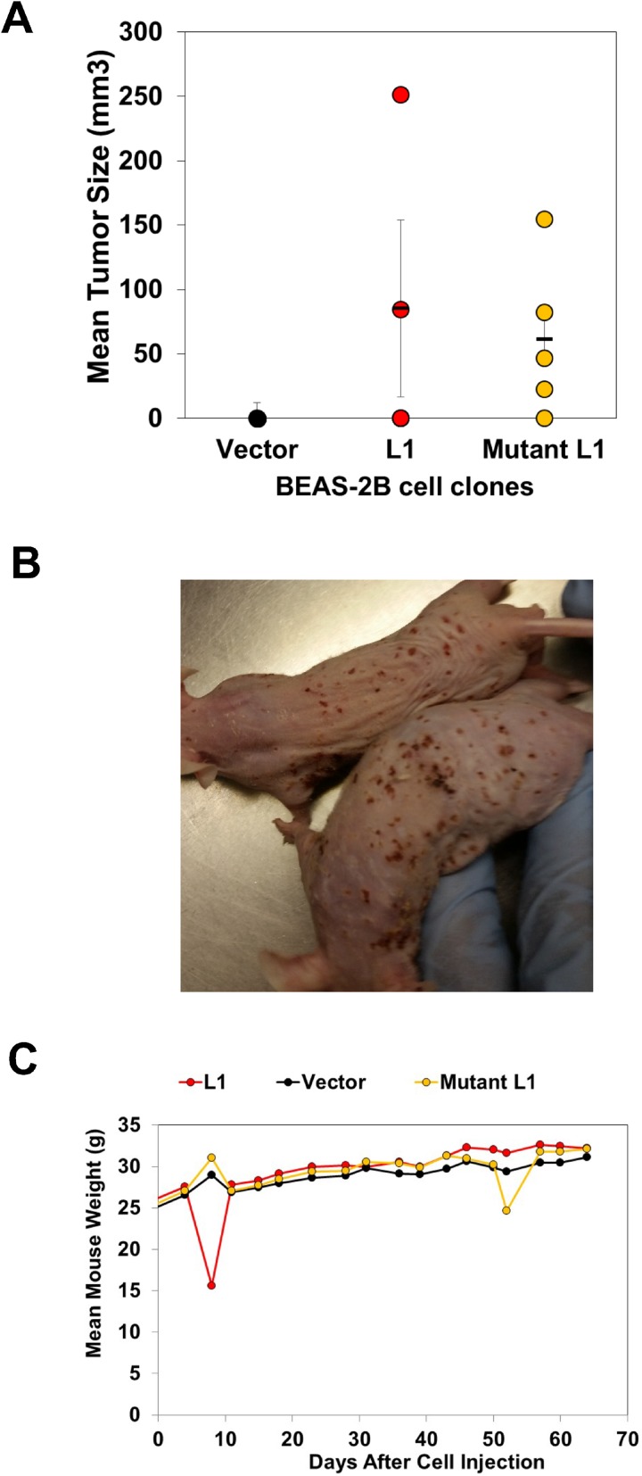
Although several lines of evidence have established the central role of epithelial-to-mesenchymal-transition (EMT) in malignant progression of non-small cell lung cancers (NSCLCs), the molecular events connecting EMT to malignancy remain poorly understood. This study presents evidence that Long Interspersed Nuclear Element-1 (LINE-1) retrotransposon couples EMT programming with malignancy in human bronchial epithelial cells (BEAS-2B). This conclusion is supported by studies showing that: 1) activation of EMT programming by TGF-β1 increases LINE-1 mRNAs and protein; 2) the lung carcinogen benzo(a)pyrene coregulates TGF-β1 and LINE-1 mRNAs, with LINE-1 positioned downstream of TGF-β1 signaling; and, 3) forced expression of LINE-1 in BEAS-2B cells recapitulates EMT programming and induces malignant phenotypes and tumorigenesis in vivo. These findings identify a TGFβ1-LINE-1 axis as a critical effector pathway that can be targeted for the development of precision therapies during malignant progression of intractable NSCLCs.

Keywords: EMT programming; LINE-1; oncogenesis.


Conflict of interest statement

CONFLICTS OF INTEREST None.

Figures

Figure 1
Figure 1. Activation of EMT Programming by TGF-β1 is Associated with LINE-1 Expression in Human Bronchial Epithelial Cells
(A) BEAS-2B whole cell lysates isolated from cells stimulated with 3 ng/mL TGF-β1 for 48 hours or control were analyzed by immunoblotting using antibodies against LINE1 (L1) ORF1 protein (ORF1p), E-cadherin, vimentin or GAPDH. (B) Total RNA from untreated or treated with 3 ng/ml TGF-β1 for 8 hours, and 1 µg of RNA was subjected to cDNA synthesis. Samples were analyzed by RT-qPCR using specific primers for human L1 (ORF1 and ORF2). Expression levels are shown as the mean of triplicates with SEM relative to controls. (C) Whole cell lysates from cells stimulated with different concentrations of TGF-β1 for 48 hours or control were analyzed for expression of L1 ORF1p by immunoblotting. Data are representative of two or more independent experiments.
Figure 2
Figure 2. Reactivation of LINE-1 by BaP is Effected via Canonical TGF-β1 Signaling
Total RNA was isolated from BEAS-2B cells treated with 0.5 uM BaP for 8 hours, and 1 µg of RNA subjected to cDNA synthesis. Samples were analyzed by RT-qPCR using specific primers for (A) human LINE-1 (ORF1 and ORF2) or (B) TGF-β1. (C) cells pre-treated with TGF-β1 receptor inhibitor (LY2157299) or vehicle (DMSO) for 30 min before BaP challenge or (D) transfected with target-specific siRNAs to SMAD2, SMAD3, or scramble siRNA or no siRNA (mock) controls. Expression levels are shown as the mean of triplicates with SEM relative to controls. (E) Whole cell extracts from transfected cells were analyzed by immunoblotting for SMAD2, SMAD3, SMAD2/3 or GAPDH antibodies (loading control) to confirm target knockdown. Data are representative of two or more independent experiments. Points represent mean of triple samples with SE.
Figure 3
Figure 3. Specificity of TGF-β1/LINE-1 Interactions in Transformed Lung Epithelial Cell Lines
NCI-H460, NCI-H520, or NCI-H1993 cell lines were challenged with BaP (0.5–2 uM) or 0.5% DMSO vehicle for 24 hours. Total RNA was isolated and 1 µg subjected to cDNA synthesis. Samples were analyzed by RT-PCR using specific primers for human TGF-β1 (A), LINE-1 (L1) ORF 1 (B) or GAPDH. Expression levels are presented relative to untreated cells. Each point represents the mean and SE of triplicate samples. The data are representative at two or more independent experiments. (C) Cells transfected with target-specific siRNAs to SNAIL or scramble siRNA or no siRNA (mock) were challenged with 3 ng/ml TGF-β1. Whole cell extracts were analyzed by immunoblotting for ORF1p, SNAIL, phospho(p)-SMAD2, or total SMAD2. Data are representative of two or more independent experiments.
Figure 4
Figure 4. Impact of LINE-1 ORF-1 siRNAs on EMT Programming
Cells were transfected with two unique target-specific siRNAs to LINE-1 targeting ORF1 regions or control siRNA (scramble) or no siRNA (mock), Forty-eight hours post-transfection, cells were challenged with 3 ng/ml TGF-β1 for an additional 48 hours or control. Whole cell lysates were analyzed by immunoblotting for ORF1p, E-Cadherin, vimentin or GAPDH.
Figure 5
Figure 5. LINE-1 Induces EMT Phenotypes in Human Bronchial Epithelial Cells
(A) Schematic representation of L1 expression vectors used to create stably transfected BEAS-2B cell clones. Vector controls lacked the L1 cassette. The mutant construct of LINE-1 carries a mutation in ORF2 (D702Y) lacking reverse transcriptase activity and rendered inactive for retrotransposition. The wild type LINE-1 construct contains retrotransposition competent ORF1 and ORF2 sequences. Both wild type and mutant vectors contain a neomycin cassette in antisense orientation to assay for retrotransposition activity. (B) Clones constitutively expressing wild type LINE-1 (L1), a mutant LINE-1 (mut L1), or empty vector control were generated by transfection using lipofectamine followed by selection of stably transfected clones with hygromycin. Whole cell lysates were analyzed by immunoblotting for L1 ORF1p, E-Cadherin, N-Cadherin, ZO1, Claudin-1, Snail-1, vimentin, or GAPDH. Data are representative two independent experiments using clones #5 (wild type L1) and #13 (mutant L1).
Figure 6
Figure 6. LINE-1 Modulates the Anti-proliferative Activity of TGF-β1 and Receptor Tyrosine Kinase Inhibitors
Clones constitutively expressing wild type L1 (L1), a mutant L1 (mut L1), or empty vector were treated with various concentrations of TGF-β1 or control (A), Receptor tyrosine kinase inhibitors: sunitinib malate (VEGFR2, PDGFRβ and c-KIT inhibitor) (B), EGFR inhibitors- Erlotinib (C) and Gefitinib (D). After 72 h of treatment, proliferation was determined by the MTT assay and normalized to untreated or DMSO (vehicle for receptor tyrosine kinase inhibitors) for each cell type. Data represent the mean plus SEM for individual samples from three independent experiments. (E) Stably transfected cells were serum-starved for 24 h and cell lysates analyzed by immunoblotting for phospho-ERK1/2 (p-Erk1/2), phospho-AKT1 (p-Akt), ERK1/2 or AKT1 (Akt). Data are representative two independent experiments using clones #5 (wild type L1) and #13 (mutant L1).
Figure 7
Figure 7. LINE-1 Induces Oncogenic Transformation of BEAS-2B cells
Clones (1 X10⁷ cells) constitutively expressing wild type LINE-1 (L1) (clone #5), a mutant L1 counterpart lacking reverse transcriptase activity (clone #13), or empty vector were mixed with matrigel and injected into 5-week old male Nu/Nu mice. (A) Tumor sizes 6-weeks after implantation. (B) Mice bearing cells expressing mutant L1. (C) Mouse weights.

References

    1. Spira A, Halmos B, Powell CA. Update in Lung Cancer 2014. Am J Resp Crit Care Med. 2015;192:283–294. - PMC - PubMed
    1. Siegel PM, Massague J. Cytostatic and apoptotic actions of TGF-beta in homeostasis and cancer. Nat Rev Cancer. 2003;3:807–821. - PubMed
    1. Reck M, Heigener DF, Mok T, Soria JC, Rabe KF. Management of non-small-cell lung cancer: recent developments. Lancet. 2013;382:709–719. - PubMed
    1. Travis WD, Brambilla E, Noguchi M, Nicholson AG, Geisinger KR, Yatabe Y, Beer DG, Powell CA, Riely GJ, Van Schil PE, Garg K, Austin JH, Asamura H, et al. International association for the study of lung cancer/american thoracic society/european respiratory society international multidisciplinary classification of lung adenocarcinoma. J Thorac Oncol. 2011;6:244–285. - PMC - PubMed
    1. Syed YY. Durvalumab: First Global Approval. Drugs. 2017;77:1369–1376. - PMC - PubMed

LinkOut - more resources