. 2017 Dec 21;17(1):261.
doi: 10.1186/s12862-017-1088-7.

Mandibulate convergence in an armoured Cambrian stem chelicerate

Affiliations

Mandibulate convergence in an armoured Cambrian stem chelicerate

Cédric Aria et al. BMC Evol Biol. .

Erratum in

Abstract

Background: Chelicerata represents a vast clade of mostly predatory arthropods united by a distinctive body plan throughout the Phanerozoic. Their origins, however, with respect to both their ancestral morphological features and their related ecologies, are still poorly understood. In particular, it remains unclear whether their major diagnostic characters were acquired early on, and their anatomical organization rapidly constrained, or if they emerged from a stem lineage encompassing an array of structural variations, based on a more labile "panchelicerate" body plan.

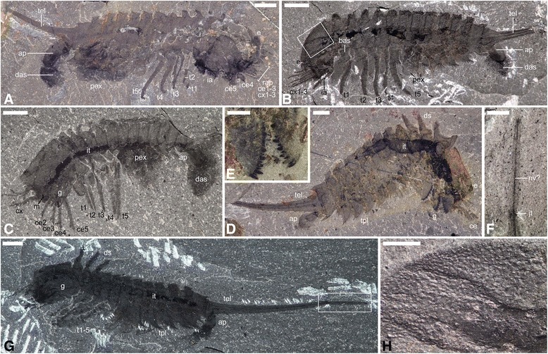
Results: In this study, we reinvestigated the problematic middle Cambrian arthropod Habelia optata Walcott from the Burgess Shale, and found that it was a close relative of Sanctacaris uncata Briggs and Collins (in Habeliida, ord. nov.), both retrieved in our Bayesian phylogeny as stem chelicerates. Habelia possesses an exoskeleton covered in numerous spines and a bipartite telson as long as the rest of the body. Segments are arranged into three tagmata. The prosoma includes a reduced appendage possibly precursor to the chelicera, raptorial endopods connected to five pairs of outstandingly large and overlapping gnathobasic basipods, antennule-like exopods seemingly dissociated from the main limb axis, and, posteriorly, a pair of appendages morphologically similar to thoracic ones. While the head configuration of habeliidans anchors a seven-segmented prosoma as the chelicerate ground pattern, the peculiar size and arrangement of gnathobases and the presence of sensory/tactile appendages also point to an early convergence with the masticatory head of mandibulates.

Conclusions: Although habeliidans illustrate the early appearance of some diagnostic chelicerate features in the evolution of euarthropods, the unique convergence of their cephalons with mandibulate anatomies suggests that these traits retained an unusual variability in these taxa. The common involvement of strong gnathal appendages across non-megacheirans Cambrian taxa also illustrates that the specialization of the head as the dedicated food-processing tagma was critical to the emergence of both lineages of extant euarthropods-Chelicerata and Mandibulata-and implies that this diversification was facilitated by the expansion of durophagous niches.

Keywords: Arthropoda; Burgess Shale; Cambrian; Chelicerata; Convergence; Macroevolution.


Conflict of interest statement

Ethics approval and consent to participate

Not applicable

Consent for publication

Not applicable

Competing interests

The authors declare that they have no competing interests.

Publisher’s Note

Springer Nature remains neutral with regard to jurisdictional claims in published maps and institutional affiliations.

Figures

Fig. 1
Fig. 1
General anatomy of Habelia optata, morphs A (d-g) and B (a-c, h). a ROMIP 64357. b USNM 139209 (inset is (h)). c ROMIP 64358. d ROMIP 64359. e Close-up of the mandibles on the counterpart of (d) (wet specimen). f Close-up of the distal telson piece in (g) (wet specimen). g Holotype USNM 57693 (inset is (f)). h Close-up on cephalic ornamentation akin to trilobite prosopon in (b). All pictures taken under cross-polarized light. For abbreviations, see Methods. Scale bars: (a), 4 mm; (b), 3 mm; (c, d, g), 2 mm; (e, f, h), 1 mm
Fig. 2
Fig. 2
Anatomical and morphological details of Habelia optata, morphs A (b, d, f, n) and B (a, e, g, i, l, m). a USNM 139209, close-up of anterior cephalic area, showing intermediary appendage. b USNM 268931, cephalon, showing superimposed insertion of endopods on gnathobases; star points to insertion of anterior endopods. c ROMIP 64357, close-up of fourth cephalic exopodial branch, distal portion showing slender podomeres; arrow points to trident of setae at podomere junction. d ROMIP 64358, close-up of anteriormost region, showing mouth opening and first anterior pairs of gnathobases. e ROMIP 64360, close-up of teeth on masticatory margin of gnathobase; note heavy concentration of carbon in teeth. f Close-up of teeth on masticatory margin of posterior gnathobase on same specimen as in D, showing stronger carbon content in dental edge. g ROMIP 64364, specimen preserved in ventral aspect, close-up of anterior region showing labrum, eyes and appendages; star marks attachment of fifth spinose endopod; arrow points at ornamental spine of cephalic pleura; insets as indicated. h ROMIP 64362, close-up of posterior trunk exopods. i ROMIP 64363, close-up of anterior right cephalic region, dorsal view showing labrum and appendages; arrows point to overprint of gnathobases underneath cephalon. j, k ROMIP 64364. j Close-up of distal portion of cephalic endopod, showing “platform” with setal brushes. k Close-up of terminal claw; arrows point to teeth on inner margin of claw. l USNM 144907, close-up of cephalic gnathobases; arrows point to dentate margins of opposing gnathobases. m ROMIP 64357, close-up on anterior left cephalic region, showing appendages; arrow points to anterior insertion of fourth cephalic endopod. n ROMIP 64359, close-up of cephalic appendages showing insertion of endopods on gnathobases; star marks attachment of fourth cephalic endopod on its gnathobase. c-f, j and k are SEM images; all other are stereomicroscope images of dry specimens under cross-polarized lighting. For abbreviations, see Methods. Scale bars: (a, g, h, i, l, n), 1 mm; (b, m), 0.5 mm; (c, d, k), 200 μm; (e), 100 μm; (f), 50 μm; (j), 500 μm
Fig. 3
Fig. 3
Diagrammatic reconstruction of Habelia optata, morph A. a Ventral view of the cephalon. Right “intermediary” appendage removed to show gnathobase morphology. b Lateral view. c Dorsal view. d Isolated biramous thoracic limb in frontal, lateral and posterior views (left to right). For abbreviations, see Methods. Line drawings courtesy of Joanna Liang © Royal Ontario Museum
Fig. 4
Fig. 4
Artistic reconstruction of Habelia optata. Courtesy of Joanna Liang © Royal Ontario Museum
Fig. 5
Fig. 5
Maximum clade credibility tree of a Bayesian analysis of arthropod relationships, using an Mkv model on a morphological matrix of 77 taxa and 215 characters. Habeliidans are in bold and red. Numbers next to nodes are posterior probabilities when <100
Fig. 6
Fig. 6
Segmental composition of major panarthropod groups as expressed through the mean of numbers of podomere (left), head limbs (center) and trunk somites (right). We use logarithmic instead of raw values to facilitate reading. Upper graphs have their error bars representing the standard deviation of the data; the error bars for lower graphs represent the minimal and maximal values inside each group. Note the punctual effect of the evolution of habeliidan heads (blue arrows) compared to the trend observed for the trunk. Tentatively, we have considered the first maxilliped in certain mandibulates as part of the functional “head,” owing to its high morphological integration to that tagma. Trilobites are not included here, their high plasticity in trunk somite number being considered autapomorphic and an arguable deviation from the general pattern seen in other artiopodans. The topology is based on phylogenetic results presented herein (Fig. 5). Double branches indicate paraphyletic groups
Fig. 7
Fig. 7
Convergences in head anatomy and morphology between Habelia (a) and selected mandibulates, in this case Ianiropsis sp. (Malacostraca: Isopoda; b; © Buz Wilson, Australian Museum) and Henicops washpoolensis (Myriapoda: Chilopoda; c; image provided by G. Edgecombe). Colours highlight the morpho-functional correspondence between sensory appendages (exopods in Habelia vs. antennae in mandibulates; green), masticatory appendages (gnathobases in Habelia vs. mandibles and maxillae in mandibulates; orange) and complimentary appendages aiding in food manipulation (seventh head appendage in Habelia vs. maxillipeds in mandibulates; blue). Note that masticatory appendages in Henicops are hidden by the large coxosternites of the maxillipeds

References

    1. Whittington HB. Early arthropods, their appendages and relationships. In: House MR, editor. The Origin of Major Invertebrate Groups. London and New York: Academic Press; 1979. pp. 253–268.
    1. Gould SJ. Wonderful life. The Burgess Shale and the nature of history. New York: Norton; 1989.
    1. Briggs DEG, Fortey RA. The early radiation and relationships of the major arthropod groups. Science. 1989;246:241–243. doi: 10.1126/science.246.4927.241. - DOI - PubMed
    1. Hou XG, Bergström J. Arthropods of the lower Cambrian Chengjiang fauna, southwest China. Fossils Strata. 1997;45:1–116.
    1. Budd GE. A palaeontological solution to the arthropod head problem. Nature. 2002;417(6886):271–275. doi: 10.1038/417271a. - DOI - PubMed