. 2018 Feb 23;200(6):e00713-17.
doi: 10.1128/JB.00713-17. Print 2018 Mar 15.

Replication Rapidly Recovers and Continues in the Presence of Hydroxyurea in Escherichia coli

Affiliations

Replication Rapidly Recovers and Continues in the Presence of Hydroxyurea in Escherichia coli

Samvel A Nazaretyan et al. J Bacteriol. .

Abstract

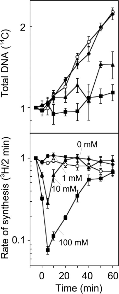
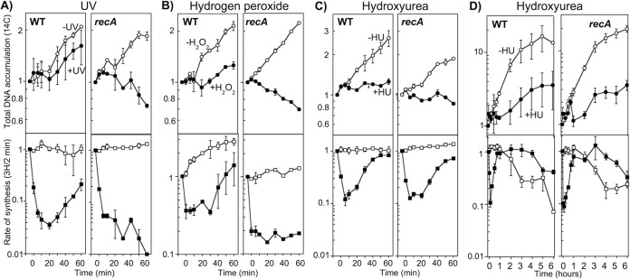
In both prokaryotes and eukaryotes, hydroxyurea is suggested to inhibit DNA replication by inactivating ribonucleotide reductase and depleting deoxyribonucleoside triphosphate pools. In this study, we show that the inhibition of replication in Escherichia coli is transient even at concentrations of 0.1 M hydroxyurea and that replication rapidly recovers and continues in its presence. The recovery of replication does not require the alternative ribonucleotide reductases NrdEF and NrdDG or the translesion DNA polymerases II (Pol II), Pol IV, and Pol V. Ribonucleotides are incorporated at higher frequencies during replication in the presence of hydroxyurea. However, they do not contribute significantly to the observed synthesis or toxicity. Hydroxyurea toxicity was observed only under conditions where the stability of hydroxyurea was compromised and by-products known to damage DNA directly were allowed to accumulate. The results demonstrate that hydroxyurea is not a direct or specific inhibitor of DNA synthesis in vivo and that the transient inhibition observed is most likely due to a general depletion of iron cofactors from enzymes when 0.1 M hydroxyurea is initially applied. Finally, the results support previous studies suggesting that hydroxyurea toxicity is mediated primarily through direct DNA damage induced by the breakdown products of hydroxyurea, rather than by inhibition of replication or depletion of deoxyribonucleotide levels in the cell.IMPORTANCE Hydroxyurea is commonly suggested to function by inhibiting DNA replication through the inactivation of ribonucleotide reductase and depleting deoxyribonucleoside triphosphate pools. Here, we show that hydroxyurea only transiently inhibits replication in Escherichia coli before replication rapidly recovers and continues in the presence of the drug. The recovery of replication does not depend on alternative ribonucleotide reductases, translesion synthesis, or RecA. Further, we show that hydroxyurea toxicity is observed only in the presence of toxic intermediates that accumulate when hydroxyurea breaks down, damage DNA, and induce lethality. The results demonstrate that hydroxyurea toxicity is mediated indirectly by the formation of DNA damage, rather than by inhibition of replication or depletion of deoxyribonucleotide levels in the cell.

Keywords: DNA replication; RNase H; hydroxyurea; translesion DNA synthesis.


Figures

FIG 1
FIG 1
DNA replication in wild-type cells is only transiently inhibited following chronic exposure to hydroxyurea. [3H]thymidine was added to [14C]thymine-prelabeled cultures for 2 min at the indicated times following treatment at time zero. The total DNA accumulation (14C) and rate of DNA synthesis (3H) relative to the amount incorporated immediately prior to exposure are plotted for wild-type cells exposed to 0 mM (circles), 1 mM (diamonds), 10 mM (triangles), or 100 mM (squares) hydroxyurea treatment. The graphs represent averages from at least three independent experiments. The error bars represent one standard error of the mean.
FIG 2
FIG 2
Cryptic class Ib and class III ribonucleotide reductases and translesion DNA polymerases do not contribute to the recovery of DNA replication in the presence of hydroxyurea. [3H]thymidine was added to [14C]thymine-prelabeled cultures for 2 min at the indicated times following treatment with 100 mM hydroxyurea (filled symbols) or mock treatment (open symbols) at time zero. The total DNA accumulation (14C; circles) and the rate of DNA synthesis (3H; squares) are plotted for wild-type (WT), nrdEF (class I ribonucleotide reductase), and nrdG (class III ribonucleotide reductase-activating enzyme) (A) and polB dinB umuDC (B) cells. Each graph represents an average from at least two independent experiments. The error bars represent one standard error of the mean.
FIG 3
FIG 3
rNTP misincorporation does not account for replication recovery in the presence of hydroxyurea. (A) Data were obtained and plotted as for Fig. 2. The total DNA accumulation (14C) in mock-treated (open circles) and hydroxyurea-treated (filled circles) cultures and the rate of DNA synthesis (3H) in mock-treated (open squares) and hydroxyurea-treated (filled squares) cultures are shown for wild-type, rnhB, and rnhA cells. The wild-type plot is reproduced from Fig. 2. (B) rNTP misincorporation during replication in the presence of hydroxyurea detectably increases in rnhB mutants. Wild-type, rnhA, and rnhB cells were exposed to 100 mM hydroxyurea and allowed to grow at 37°C. At the indicated times, genomic DNA was purified and analyzed on alkali- and neutral-agarose gels. Representative gels are shown. Lanes M, lambda HindIII size marker; lanes U, untreated cells. (C) [3H]thymidine was added to [14C]thymine-prelabeled wild-type cultures for 2 min at the indicated times following treatment with 100 mM hydroxyurea (filled symbols) or mock treatment (open symbols) at time zero. Samples were then lysed in the presence or absence of 500 mM NaOH. The total DNA accumulation (14C; circles) and rate of DNA synthesis (3H; squares) are plotted. The graphs represent averages from at least two independent experiments. The error bars represent one standard error of the mean.
FIG 4
FIG 4
RecA is not required for replication recovery following treatment with hydroxyurea, suggesting an absence of DNA damage. (A) [3H]thymidine was added to [14C]thymine-prelabeled cultures for 2 min at the indicated times following 27 J/m2 UV irradiation (filled symbols) or mock irradiation (open symbols) at time zero. The total DNA accumulation (14C; circles) and rate of DNA synthesis (3H; squares) are plotted. (B) Cells were either exposed to 10 mM hydrogen peroxide for 5 min (filled symbols) or mock treated (open symbols) at time zero and then allowed to recover in the presence of 200 μg/ml catalase. [3H]thymidine was added to [14C]thymine-prelabeled cultures for 2 min at the indicated times following treatment. The total DNA accumulation (14C; circles) and rate of DNA synthesis (3H; squares) are plotted. (C) Data were obtained and plotted as for Fig. 2. The total DNA accumulations (14C) in mock-treated (open circles) and hydroxyurea-treated (filled circles) cultures and rates of DNA synthesis (3H) in mock-treated (open squares) and hydroxyurea-treated (filled squares) cultures are shown. The wild-type plot is reproduced from Fig. 2. (D) Data were obtained and plotted as for Fig. 2. The total DNA accumulations (14C) in mock-treated (open circles) and hydroxyurea-treated (filled circles) cultures and rates of DNA synthesis (3H) in mock-treated (open squares) and hydroxyurea-treated (filled squares) cultures are shown. All the graphs represent averages from at least two independent experiments. The error bars represent one standard error of the mean.
FIG 5
FIG 5
RecA is required for cell survival and replication recovery following treatment with heat-decayed hydroxyurea, consistent with the induction of DNA damage by toxic by-products of hydroxyurea. (A) Survival of wild-type and recA cells on agar plates supplemented with 10 mM hydroxyurea. (B) The survival of wild-type (squares) and recA (circles) cells after exposure to 100 mM heat-decayed (HU+heat; closed symbols) or freshly prepared (HU; open symbols) hydroxyurea is plotted following treatment in liquid cultures for the indicated times. (C) [3H]thymidine was added to [14C]thymine-prelabeled cultures for 2 min at the indicated times following treatment with heat-decayed 100 mM hydroxyurea (filled symbols) or mock treatment (open symbols) at time zero. The total DNA accumulation (14C; circles) and rate of DNA synthesis (3H; squares) are plotted. All the graphs represent an average of at least two independent experiments. The error bars represent one standard error of the mean. (D) RecA, but not the cryptic class Ib and class III ribonucleotide reductases, translesion DNA polymerases, or ribonucleases HI and HII, is hypersensitive to hydroxyurea in plates. The survival of wild-type parental (squares), nrdEF (inverted triangles), nrdG (diamonds), polB dinB umuDC (open circles), rnhA (open triangles), rnhB (closed triangles), and recA (closed circles) cultures is plotted following growth on hydroxyurea-containing agar plates at the indicated concentrations. The graphs represent averages from at least two independent experiments. The error bars represent one standard error of the mean.
FIG 6
FIG 6
Model depicting the proposed effects of hydroxyurea, or heat-degraded hydroxyurea, on cellular metabolism.

References

    1. Stearns B, Losee KA, Bernstein J. 1963. Hydroxyurea. A new type of potential antitumor agent. J Med Chem 6:201. - PubMed
    1. Stauber RH, Knauer SK, Habtemichael N, Bier C, Unruhe B, Weisheit S, Spange S, Nonnenmacher F, Fetz V, Ginter T, Reichardt S, Liebmann C, Schneider G, Krämer OH. 2012. A combination of a ribonucleotide reductase inhibitor and histone deacetylase inhibitors downregulates EGFR and triggers BIM-dependent apoptosis in head and neck cancer. Oncotarget 3:31–43. doi:10.18632/oncotarget.430. - DOI - PMC - PubMed
    1. Piccinini G, Foli A, Comolli G, Lisziewicz J, Lori F. 2002. Complementary antiviral efficacy of hydroxyurea and protease inhibitors in human immunodeficiency virus-infected dendritic cells and lymphocytes. J Virol 76:2274–2278. doi:10.1128/jvi.76.5.2274-2278.2002. - DOI - PMC - PubMed
    1. Segal JB, Strouse JJ, Beach MC, Haywood C, Witkop C, Park H, Wilson RF, Bass EB, Lanzkron S. 2008. Hydroxyurea for the treatment of sickle cell disease. Evid Rep Technol Assess (Full Rep) 165:1–95. - PMC - PubMed
    1. Elford HL. 1968. Effect of hydroxyurea on ribonucleotide reductase. Biochem Biophys Res Commun 33:129–135. doi:10.1016/0006-291X(68)90266-0. - DOI - PubMed

Publication types

LinkOut - more resources