. 2018 Feb 20;86(3):e00615-17.
doi: 10.1128/IAI.00615-17. Print 2018 Mar.

Immunoproteasome Subunits Are Required for CD8+ T Cell Function and Host Resistance to Brucella abortus Infection in Mice

Affiliations

Immunoproteasome Subunits Are Required for CD8+ T Cell Function and Host Resistance to Brucella abortus Infection in Mice

Gabriela Guimarães et al. Infect Immun. .

Abstract

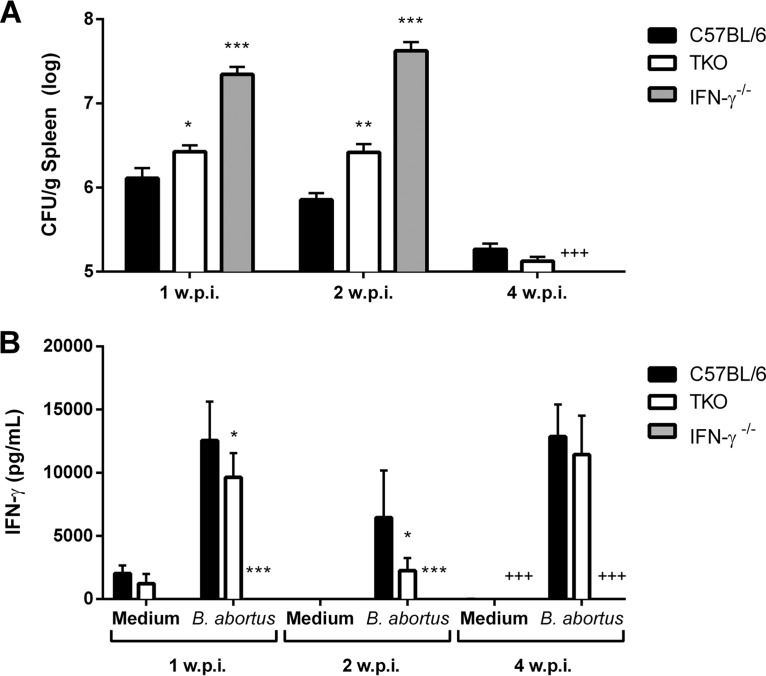
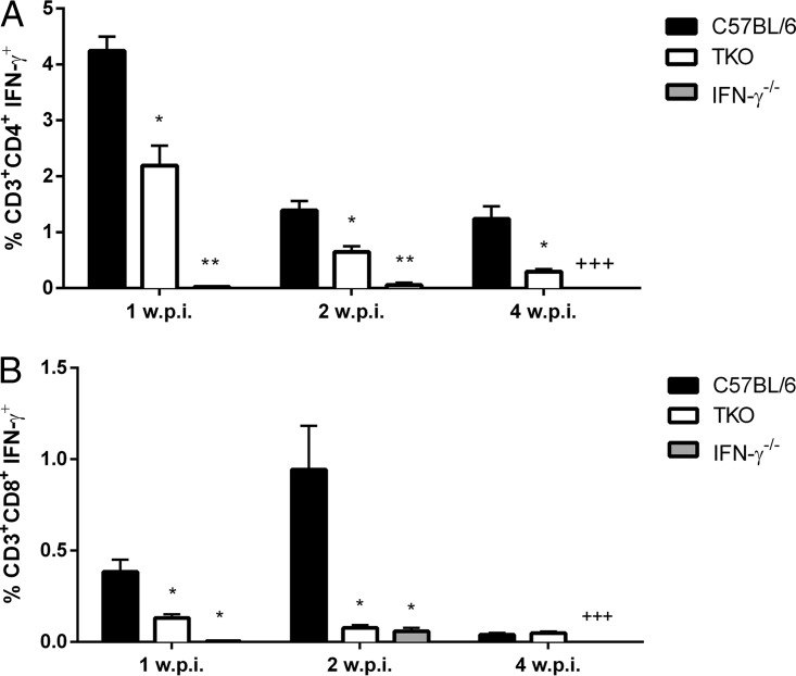
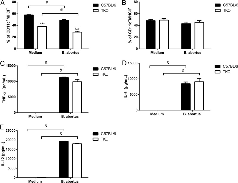
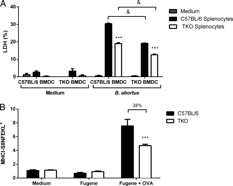
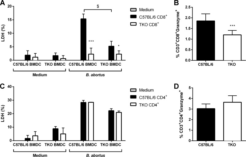
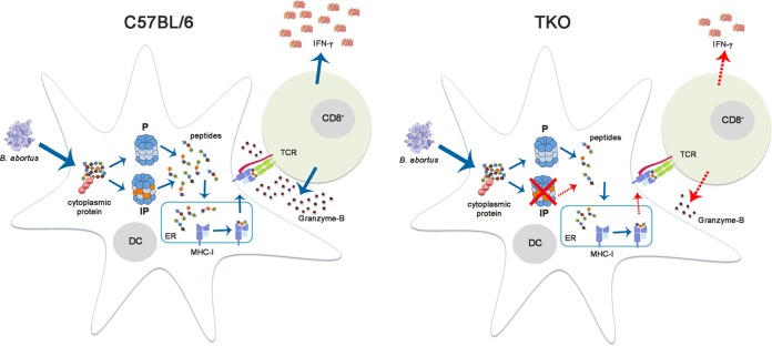
The immunoproteasome is a specific proteasome isoform composed of three subunits, termed β1i, β2i, and β5i. Its proteolytic activity enhances the quantity and quality of peptides to be presented by major histocompatibility complex class I (MHC-I) molecules to CD8+ T cells. However, the role of the combined deficiency of the three immunoproteasome subunits in protective immunity against bacterial pathogens has not been investigated. In this study, we addressed the role of the immunoproteasome during infection by Brucella abortus, an intracellular bacterium that requires CD8+ T cell responses for the control of infection. Here, we demonstrate that immunoproteasome triple-knockout (TKO) mice were more susceptible to Brucella infection. This observed susceptibility was accompanied by reduced interferon gamma (IFN-γ) production by mouse CD4+ and CD8+ T lymphocytes. Moreover, the absence of the immunoproteasome had an impact on MHC-I surface expression and antigen presentation by dendritic cells. CD8+ T cell function, which plays a pivotal role in B. abortus immunity, also presented a partial impairment of granzyme B expression and, consequently, reduced cytotoxic activity. In conclusion, these results strongly suggest that immunoproteasome subunits are important components in host resistance to B. abortus infection by impacting both the magnitude and quality of CD8+ T cell responses.

Keywords: Brucella abortus; CD8+ T cells; MHC-I; immunoproteasome.


Figures

FIG 1
FIG 1
Lack of the immunoproteasome renders mice susceptible to B. abortus infection. C57BL/6, TKO, and IFN-γ−/− mice were intraperitoneally inoculated with 106 CFU of B. abortus S2308. (A) The number of bacteria in the spleen was analyzed by counting of CFU after 1, 2, and 4 weeks of infection. (B) At 1, 2, or 4 weeks postinfection (w.p.i.), spleen cells (1 × 10⁶ cells) from C57BL/6, TKO, and IFN-γ−/− were restimulated with B. abortus S2308 (MOI of 100:1), and IFN-γ levels were measured by an ELISA after 72 h. +++ indicates death of animals. Data are the means ± standard deviations of results for five mice/group and are representative of data from three independent experiments. *, P < 0.05; **, P < 0.01; ***, P < 0.001 (for statistically significant differences compared to C57BL/6 mice).
FIG 2
FIG 2
The immunoproteasome is critical for IFN-γ production by CD4+ and CD8+ T cells after B. abortus infection. Flow cytometry analysis of C57BL/6, TKO, and IFN-γ−/− splenocytes obtained after 1, 2, and 4 weeks of infection with B. abortus was performed after 4 h of incubation with brefeldin A and concanavalin A. Cells were assessed for CD3+ CD4+ IFN-γ+ (A) and CD3+ CD8+ IFN-γ+ (B) populations. A total of 100,000 events was obtained and analyzed. +++ indicates death of animals. Data are the means ± standard deviations of results for five mice/group and are representative of data from three independent experiments. *, P < 0.05; **, P < 0.01 (for statistically significant differences compared to C57BL/6 mice).
FIG 3
FIG 3
MHC class I surface expression on dendritic cells from TKO animals is partially impaired. Flow cytometry analysis of C57BL/6 and TKO mouse splenocytes was performed at the second week of infection with B. abortus. (A and B) Cells were assessed for CD11c+ MHC-I+ (A) and CD11c+ MHC-II+ (B) populations. A total of 100,000 events was obtained and analyzed. Dendritic cells were cultured (5 × 10⁵ cells/well) and stimulated with B. abortus S2308 (MOI of 100:1) for 24 h. (C to E) Culture supernatants were harvested, and levels of TNF-α (C), IL-6 (D), and IL-12 (E) secretion were determined by an ELISA according to the manufacturer's instructions. Data are the means ± standard deviations of results from five mice/group and are representative of data from three independent experiments. ***, P < 0.001 (for statistically significant differences compared to C57BL/6 mice). #, P < 0.01; &, P < 0.001 (for statistically significant differences between Brucella-infected and uninfected cells).
FIG 4
FIG 4
Antigen presentation by Brucella-infected TKO dendritic cells is partially impaired. (A) Differentiated dendritic cells (5 × 10⁵ cells/well) obtained from C57BL/6 and TKO mice were infected with B. abortus (MOI of 100:1) and used as target cells. Splenocytes (1 × 10⁶ cells/well) obtained from C57BL/6 and TKO mice at the second week of infection were used as effector cells for cytotoxicity assays and were cocultured with dendritic cells in 24-well plates in DMEM. Effector cells were added to target cells in duplicate at a 2:1 ratio, and 24 h after coculture, the supernatant was collected to quantify LDH release according to the manufacturer's instructions. (B) Dendritic cells from C57BL/6 and TKO mice were incubated with OVA-Fugene for 2 h. Cells were then stained with anti-SIINFEKL–H-2b and analyzed by flow cytometry. A total of 100,000 events was obtained and analyzed. Data are the means ± standard deviations of results for five mice/group and are representative of data from three independent experiments. ***, P < 0.001 (for statistically significant differences compared to C57BL/6 mice); &, P < 0.001 (for statistically significant differences between C57BL/6 and TKO dendritic cells cocultured with C57BL/6 or TKO splenocytes).
FIG 5
FIG 5
TKO CD8+ T cell cytotoxic activity is reduced in B. abortus-infected mice. Bone marrow-derived dendritic cells (5 × 10⁵ cells/well) obtained from C57BL/6 and TKO mice were infected with B. abortus (MOI of 100:1) and used as target cells. (A and C) CD8+ (A) or CD4+ (C) T lymphocytes (1 × 10⁶ cells/well) obtained by cell sorting from C57BL/6 and TKO splenocytes at the second week of infection were used as effector cells for cytotoxicity assays and were cocultured with dendritic cells in 24-well plates in DMEM. CD8+ T cells were added to target cells at a 1:2 ratio, CD4+ T cells were added to target cells in duplicate at a 1:1 ratio, and 24 h after coculture, the supernatant was collected to quantify LDH release according to the manufacturer's instructions. (B and D) Percentages of CD3+ CD8+ granzyme B-positive (B) and CD3+ CD4+ granzyme B-positive (D) cells measured by flow cytometry in splenocytes from C57BL/6 and TKO mice. Data are the means ± standard deviations of results for five mice/group and are representative of data from three independent experiments. *, P < 0.05; ***, P < 0.001 (for statistically significant differences compared to C57BL/6 mice). $, P < 0.05 (for statistically significant differences between wild-type and TKO dendritic cells cocultured with C57BL/6 CD8+ T cells).
FIG 6
FIG 6
Schematic model of the CD8+ T cell response during B. abortus infection in TKO animals. B. abortus proteins secreted into the cytoplasm of dendritic cells are degraded by the immunoproteasome, generating a variety of peptides. These peptides are then transported to the endoplasmic reticulum and bind to the MHC-I molecule. The MHC-I–peptide complex is exported to the cell membrane and recognized by CD8+ T lymphocytes. After this, CD8+ T cells produce granzyme B and IFN-γ in response to infection. The red dotted arrows indicate the partially impaired process in TKO dendritic and CD8+ T cells. P, proteasome; IP, immunoproteasome; ER, endoplasmic reticulum; DC, dendritic cell.

Similar articles

Cited by

References

    1. Russell JH, Ley TJ. 2002. Lymphocyte-mediated cytotoxicity. Annu Rev Immunol 20:323–370. doi:10.1146/annurev.immunol.20.100201.131730. - DOI - PubMed
    1. Kagi D, Vignaux F, Ledermann B, Burki K, Depraetere V, Nagata S, Hengartner H, Golstein P. 1994. Fas and perforin pathways as major mechanisms of T cell-mediated cytotoxicity. Science 265:528–530. doi:10.1126/science.7518614. - DOI - PubMed
    1. Janeway CA., Jr 1992. The T cell receptor as a multicomponent signalling machine: CD4/CD8 coreceptors and CD45 in T cell activation. Annu Rev Immunol 10:645–674. doi:10.1146/annurev.iy.10.040192.003241. - DOI - PubMed
    1. Kloetzel P-M, Ossendorp F. 2004. Proteasome and peptidase function in MHC-class-I-mediated antigen presentation. Curr Opin Immunol 16:76–81. doi:10.1016/j.coi.2003.11.004. - DOI - PubMed
    1. Lowe J, Stock D, Jap B, Zwickl P, Baumeister W, Huber R. 1995. Crystal structure of the 20S proteasome from the archaeon T. acidophilum at 3.4 A resolution. Science 268:533–539. doi:10.1126/science.7725097. - DOI - PubMed

Publication types

MeSH terms

LinkOut - more resources