Comparative Study
. 2018 Feb 20;86(3):e00812-17.
doi: 10.1128/IAI.00812-17. Print 2018 Mar.

Autoimmune-Disease-Prone NOD Mice Help To Reveal a New Genetic Locus for Reducing Pulmonary Disease Caused by Mycoplasma pulmonis

Affiliations
Comparative Study

Autoimmune-Disease-Prone NOD Mice Help To Reveal a New Genetic Locus for Reducing Pulmonary Disease Caused by Mycoplasma pulmonis

Nadeeka K Wawegama et al. Infect Immun. .

Abstract

Mycoplasmas are bacterial pathogens of a range of animals, including humans, and are a common cause of respiratory disease. However, the host genetic factors that affect resistance to infection or regulate the resulting pulmonary inflammation are not well defined. We and others have previously demonstrated that nonobese diabetic (NOD) mice can be used to investigate disease loci that affect bacterial infection and autoimmune diabetes. Here we show that NOD mice are more susceptible than C57BL/6 (B6) mice to infection with Mycoplasma pulmonis, a natural model of pulmonary mycoplasmosis. The lungs of infected NOD mice had higher loads of M. pulmonis and more severe inflammatory lesions. Moreover, congenic NOD mice that harbored different B6-derived chromosomal intervals enabled identification and localization of a new mycoplasmosis locus, termed Mpr2, on chromosome 13. These congenic NOD mice demonstrated that the B6 allele for Mpr2 reduced the severity of pulmonary inflammation caused by infection with M. pulmonis and that this was associated with altered cytokine and chemokine concentrations in the infected lungs. Mpr2 also colocalizes to the same genomic interval as Listr2 and Idd14, genetic loci linked to listeriosis resistance and autoimmune diabetes susceptibility, respectively, suggesting that allelic variation within these loci may affect the development of both infectious and autoimmune disease.

Keywords: Mycoplasma pulmonis; autoimmune disease; complex genetic trait; congenic mice; infectious disease; murine respiratory mycoplasmosis; nonobese diabetic mouse; pulmonary inflammation; resistance loci.


Figures

FIG 1
FIG 1
Pulmonary mycoplasma load and pathology in mice at 3 days postinfection. NOD, B6, and congenic NOD mice were infected with 4 × 10⁷ CCU of M. pulmonis at approximately 10 weeks of age. No mice were diabetic prior to or during the course of infection. Mycoplasma load was determined at 3 days postinfection in the right lung (A), and the total lung lesion index was determined by histopathological examination of the left lung (B). The three lung lesion scores that make up the total lung lesion index are presented separately: severity of lymphoid infiltration (C), extent of airway exudate (D), and extent of alveolar exudate (E). The limit of detection was 50 CCU/ml; lung homogenates below this limit were estimated to contain 50 CCU/ml (log10 = 1.7) for the purposes of data presentation and statistical analysis. The horizontal lines indicate the grand means, determined by combining results for females and males. Asterisks indicate significance (*, P ≤ 0.05; **, P ≤ 0.01). There was no significant difference between females and males within strains for either mycoplasma load or total lung lesion index.
FIG 2
FIG 2
Pulmonary mycoplasma load and pathology in mice at 7 days postinfection. NOD, B6, and congenic NOD mice were infected with M. pulmonis at approximately 10 weeks of age. Data in all panels represent the combination of two independent experiments (for experiment 1, n = 5 or 6 males and 5 to 8 females per strain, infected with 1.7 × 10⁷ CCU; for experiment 2, n = 8 males and 8 females per strain, infected with 6.2 × 10⁷ CCU). No mice were diabetic prior to or during the course of infection. Mycoplasma load was determined at 7 days postinfection in the right lung (A), and total lung lesion index was determined by histopathological examination of the left lung (B). The three lung lesion scores that make up the total lung lesion index are presented separately: severity of lymphoid infiltration (C), extent of airway exudate (D), and extent of alveolar exudate (E). The limit of accurate detection was 50 CCU/ml; lung homogenates below this limit were estimated to contain 50 CCU/ml (log10 = 1.7) for the purposes of data presentation and statistical analysis. The horizontal lines indicate the grand means, determined by combining results for females and males. Asterisks indicate significance (*, P ≤ 0.05; **, P ≤ 0.01; ***, P ≤ 0.001; ****, P ≤ 0.0001). There was no significant difference between females and males within strains for either mycoplasma load or total lung lesion index.
FIG 3
FIG 3
Pulmonary mycoplasma load and pathology in mice at 14 days postinfection. NOD, B6, and congenic NOD mice were infected with 4 × 10⁷ CCU of M. pulmonis at approximately 10 weeks of age. No mice were diabetic prior to or during the course of infection. Mycoplasma load was determined at 14 days postinfection in the right lung (A), and total lung lesion index was determined by histopathological examination of the left lung (B). The three lung lesion scores that make up the total lung lesion index are presented separately: severity of lymphoid infiltration (C), extent of airway exudate (D), and extent of alveolar exudate (E). The limit of accurate detection was 50 CCU/ml; lung homogenates below this limit were estimated to contain 50 CCU/ml (log10 = 1.7) for the purposes of data presentation and statistical analysis. The horizontal lines indicate the grand means, determined by combining results for females and males. Asterisks indicate significance (*, P ≤ 0.05; **, P ≤ 0.01; ***, P ≤ 0.001; ****, P ≤ 0.0001). There was no significant difference between females and males within strains for either mycoplasma load or total lung lesion index.
FIG 4
FIG 4
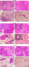
Lungs from infected NOD and R6 mice have more severe pulmonary pathology. Representative histopathological changes are shown for H&E-stained left lungs from infected mice at 3, 7, and 14 days postinfection. The majority of lungs from infected B6 mice showed healthy parenchyma with little to no peribronchiolar or perivascular infiltration of lymphocytes or influx of polymorphonuclear cells into the bronchial lumen or parenchyma at all three time points postinfection. In general, lungs from infected R8 mice showed unorganized lymphocytes surrounding the bronchi, with the lung parenchyma and bronchial lumen free of polymorphonuclear cells at 3 days postinfection. At 7 and 14 days postinfection, the majority of lungs from R8 mice had a few layers of organized lymphocytes surrounding the bronchi, with only patches of polymorphonuclear cells in the parenchyma. In contrast, the majority of lungs from infected NOD and R6 mice had extensive lymphocytic infiltration at all time points postinfection. This infiltration appeared as organized layers around the bronchi and blood vessels, with bronchi filled with polymorphonuclear cells and lymphocytes (e.g., day 7 R6 inset) and alveoli having extensive infiltration of polymorphonuclear cells (e.g., day 7 NOD inset). A, alveoli; B, bronchi; E, bronchial epithelium; L, lymphoid aggregation; P, polymorphonuclear cells; V, blood vessels. Bars = 50 μm, except for day 7 insets (NOD and R6, bar = 5 μm; R8, bar = 20 μm; and B6, bar = 10 μm).
FIG 5
FIG 5
Proinflammatory cytokines IL-6, TNF-α, and IFN-γ are detected in lungs infected with M. pulmonis. Homogenates were prepared from the lungs of female and male mice at 3 days after infection with 4 × 10⁷ CCU of M. pulmonis and used to measure the levels of IFN-γ (A), TNF-α (D), IL-6 (G), and IL-4 (J). Homogenates were prepared from the lungs of female and male mice at 7 days after infection with 6.2 × 10⁷ CCU of M. pulmonis and used to measure the levels of IFN-γ (B), TNF-α (E), IL-6 (H), and IL-4 (K). Homogenates were prepared from the lungs of female and male mice at 14 days after infection with 4 × 10⁷ CCU of M. pulmonis and used to measure the levels of IFN-γ (C), TNF-α (F), IL-6 (I), and IL-4 (L). For day 3 and day 14, the BD Biosciences cytometric bead array was used; the limit of detection of IFN-γ was 0.5 pg/ml (log2 = −1), that of TNF-α was 0.9 pg/ml (log2 = −0.15), that of IL-6 was 1.4 pg/ml (log2 = 0.49), and that of IL-4 was 0.03 pg/ml (log2 = −5.1). For day 7, the eBiosciences cytokine bead array was used; the limit of detection of IFN-γ was 6.5 pg/ml (log2 = 2.7), that of TNF-α was 2.1 pg/ml (log2 = 1.07), that of IL-6 was 2.2 pg/ml (log2 = 1.14), and that of IL-4 was 0.7 pg/ml (log2 = −0.51). Samples with concentrations below these thresholds were assumed to have concentrations equal to the limit of detection of the assay for the purposes of statistical analysis. The horizontal lines indicate the grand means, determined by combining results for females and males. Asterisks indicate significance (*, P ≤ 0.05; **, P ≤ 0.01; ***, P ≤ 0.001).
FIG 6
FIG 6
Chemokines MCP2/CCL8 and MIP-1β/CCL4 are detected in lungs infected with M. pulmonis. Homogenates were prepared from the lungs of NOD, R6, R8, and B6 mice at 7 days after infection with 6.2 × 10⁷ CCU and NOD and B6 mice at 14 days after infection with 4 × 10⁷ CCU of M. pulmonis. MCP2/CCL8 (A) and MIP-1β/CCL4 (C) were measured in NOD and B6 mice at both 7 and 14 days postinfection. MCP2/CCL8 (B) and MIP-1β/CCL4 (D) were also measured in lung homogenates at 7 days postinfection in R6 and R8 mice (NOD and B6 results for 7 days postinfection shown again for comparison). The limit of accurate detection of MCP2 was 6.25 pg/ml (log2 = 2.64) and that of MIP-1α was 12.5 pg/ml (log2 = 3.64); samples with concentrations below these thresholds were assumed to have concentrations equal to the limit of detection of the assay for the purposes of statistical analysis. The horizontal lines indicate the grand means, determined by combining results for females and males. Asterisks indicate significance (*, P ≤ 0.05; **, P ≤ 0.01; ***, P ≤ 0.001; ****, P ≤ 0.0001).

References

    1. Lindsey JR, Baker HJ, Overcash RG, Cassell GH, Hunt CE. 1971. Murine chronic respiratory disease. Significance as a research complication and experimental production with Mycoplasma pulmonis. Am J Pathol 64:675–708. - PMC - PubMed
    1. Cassell GH, Lindsey JR, Davis JK, Davidson MK, Brown MB, Mayo JG. 1981. Detection of natural Mycoplasma pulmonis infection in rats and mice by an enzyme linked immunosorbent assay (ELISA). Lab Anim Sci 31:676–682. - PubMed
    1. Cassell GH. 1982. Derrick Edward Award Lecture. The pathogenic potential of mycoplasmas: Mycoplasma pulmonis as a model. Rev Infect Dis 4(Suppl):S18–S34. - PubMed
    1. Cartner SC, Lindsey JR, Gibbs-Erwin J, Cassell GH, Simecka JW. 1998. Roles of innate and adaptive immunity in respiratory mycoplasmosis. Infect Immun 66:3485–3491. - PMC - PubMed
    1. Lai WC, Bennett M, Pakes SP, Kumar V, Steutermann D, Owusu I, Mikhael A. 1990. Resistance to Mycoplasma pulmonis mediated by activated natural killer cells. J Infect Dis 161:1269–1275. doi:10.1093/infdis/161.6.1269. - DOI - PubMed

Publication types

LinkOut - more resources