Review
. 2017 Dec 20;26(146):170025.
doi: 10.1183/16000617.0025-2017. Print 2017 Dec 31.

Lung nodules: size still matters

Affiliations
Review

Lung nodules: size still matters

Anna Rita Larici et al. Eur Respir Rev. .

Abstract

The incidence of indeterminate pulmonary nodules has risen constantly over the past few years. Determination of lung nodule malignancy is pivotal, because the early diagnosis of lung cancer could lead to a definitive intervention. According to the current international guidelines, size and growth rate represent the main indicators to determine the nature of a pulmonary nodule. However, there are some limitations in evaluating and characterising nodules when only their dimensions are taken into account. There is no single method for measuring nodules, and intrinsic errors, which can determine variations in nodule measurement and in growth assessment, do exist when performing measurements either manually or with automated or semi-automated methods. When considering subsolid nodules the presence and size of a solid component is the major determinant of malignancy and nodule management, as reported in the latest guidelines. Nevertheless, other nodule morphological characteristics have been associated with an increased risk of malignancy. In addition, the clinical context should not be overlooked in determining the probability of malignancy. Predictive models have been proposed as a potential means to overcome the limitations of a sized-based assessment of the malignancy risk for indeterminate pulmonary nodules.


Conflict of interest statement

Conflict of interest: None declared.

Figures

FIGURE 1
FIGURE 1
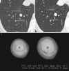
Limitations of two-dimensional (2D) measurements. The axial diameter may not be the maximum one in the evaluation of lung nodules. a) A small part-solid nodule in the apico-posterior segment of the left upper lobe, with a maximum axial diameter of 12×12.2 mm; b) the sagittal multiplanar reconstruction shows that the largest diameter of the same nodule is the sagittal one of 24.7 mm. The multiplanar evaluation of nodule diameter is especially important to document asymmetrical growth of nodules. c), d) The low level of agreement when measuring small nodules: for the same nodule in the right lower lobe two different diameter values have been reported by two readers. Considering the nearest whole diameter of the two values, it results in 1 mm difference in the maximum diameter, a significant difference when considering small nodules.
FIGURE 2
FIGURE 2
Disagreement in measuring the solid portion of a part-solid nodule when using different reconstruction algorithms and window settings. A part-solid nodule in the apical segment of left lower lobe is shown. a) By using a high-spatial frequency algorithm and the lung window, the measured maximum axial diameter of the solid portion of the nodule corresponds to 20.3 mm; b) by using a smooth algorithm and the mediastinal window, the measured maximum axial diameter of the solid portion of the nodule corresponds to 16 mm. 2D: two-dimensional.
FIGURE 3
FIGURE 3
Volume evaluation during follow-up allows the detection of nodule growth over a shorter period of time compared to diameter estimation. a) Computed tomography (CT) axial image shows the same nodule located in the right lower lobe as reported in figure 1c; b) a 3-month follow-up axial CT image demonstrates minimal change in nodule diameters; c) conversely, nodule volume calculation using a three-dimensional (3D) volumetric method demonstrates a significant increase in volume within the range of malignancy. Histopathology revealed a carcinoid tumour. 2D: two-dimensional; TV: total volume; DT: volume doubling time; %G: volume increase; scan inter: scan interval. Squares in the nodule represent the starting points of the 3D analysis.

Comment in

  • doi: 10.1183/16000617.0049-2017

References

    1. Hansell DM, Bankier AA, MacMahon H, et al. Fleischner Society: glossary of terms for thoracic imaging. Radiology 2008; 246: 697–722. - PubMed
    1. Callister ME, Baldwin DR, Akram AR, et al. British Thoracic Society guidelines for the investigation and management of pulmonary nodules. Thorax 2015; 70: Suppl. ii1–ii54. - PubMed
    1. Swensen SJ, Silverstein MD, Ilstrup DM, et al. The probability of malignancy in solitary pulmonary nodules. Application to small radiologically indeterminate nodules. Arch Intern Med 1997; 157: 849–855. - PubMed
    1. MacMahon H, Austin JH, Gamsu G, et al. Guidelines for management of small pulmonary nodules detected on CT scans: a statement from the Fleischner Society. Radiology 2005; 237: 395–400. - PubMed
    1. Gould MK, Donington J, Lynch WR, et al. Evaluation of individuals with pulmonary nodules: when is it lung cancer? Diagnosis and management of lung cancer, 3rd ed: American College of Chest Physicians evidence-based clinical practice guidelines. Chest 2013; 143: Suppl. e93S–e120S. - PMC - PubMed

MeSH terms