Review
. 2017 Dec 20;26(146):170094.
doi: 10.1183/16000617.0094-2017. Print 2017 Dec 31.

Metabolic dysfunction in pulmonary hypertension: from basic science to clinical practice

Affiliations
Review

Metabolic dysfunction in pulmonary hypertension: from basic science to clinical practice

Stephen Y Chan et al. Eur Respir Rev. .

Erratum in

Abstract

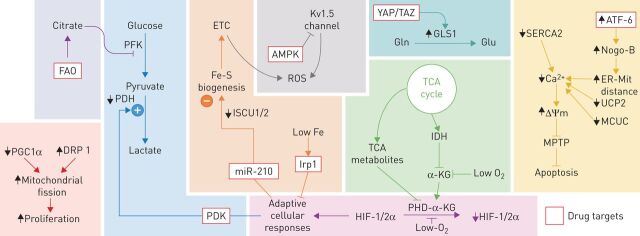
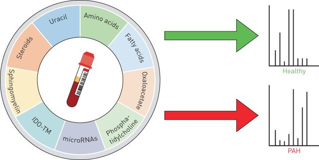
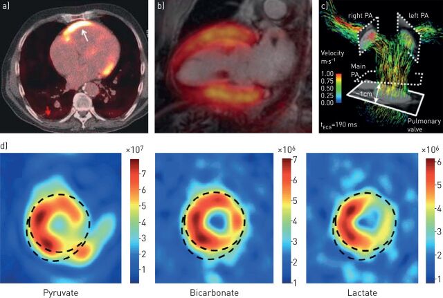
Pulmonary hypertension (PH) is an often-fatal vascular disease of unclear molecular origins. The pulmonary vascular remodelling which occurs in PH is characterised by elevated vasomotor tone and a pro-proliferative state, ultimately leading to right ventricular dysfunction and heart failure. Guided in many respects by prior evidence from cancer biology, recent investigations have identified metabolic aberrations as crucial components of the disease process in both the pulmonary vessels and the right ventricle. Given the need for improved diagnostic and therapeutic options for PH, the development or repurposing of metabolic tracers and medications could provide an effective avenue for preventing or even reversing disease progression. In this review, we describe the metabolic mechanisms that are known to be dysregulated in PH; we explore the advancing diagnostic testing and imaging modalities that are being developed to improve diagnostic capability for this disease; and we discuss emerging drugs for PH which target these metabolic pathways.


Conflict of interest statement

Conflict of interest: Disclosures can be found alongside this article at err.ersjournals.com

Figures

FIGURE 1
FIGURE 1
Overview of the dysfunctional metabolic pathways in pulmonary vascular cell types implicated in the development of pulmonary hypertension. KG: ketoglutarate; ΔΨm: mitochondrial membrane potential; AMPK: adenosine monophosphate-activated protein kinase; ATF: activating transcription factor; Ca2+: calcium; DRP: dynamin-related protein; ER: endoplasmic reticulum; ETC: electron transport chain; FAO: fatty acid oxidation; Fe: iron; S: sulfur; Gln: glutamine; GLS1: glutaminase; Glu: glutamate; HIF: hypoxia-inducible factor; IDH: isocitrate dehydrogenase; Irp: iron-regulatory protein; ISCU: iron-sulfur cluster assembly protein; Kv1.2: voltage-dependent potassium channel 1.2; MCUC: mitochondrial calcium uniporter complex; miR-210: micro-RNA 210; Mit: mitochondria; MPTP: mitochondrial permeability transition pore; Nogo-B: neurite outgrowth inhibitor-B; O2: oxygen: PDH: pyruvate dehydrogenase; PDK: pyruvate dehydrogenase kinase; PFK: phosphofructokinase; PGC: proliferator-activated receptor-y coactivator; PHD: prolyl hydroxlase; ROS: reactive oxygen species; SERCA: sarco-/endoplasmic reticulum calcium-ATPase; TAZ: transcription coactivator with a PDZ-binding motif; TCA: tricarboxylic acid; UCP: uncoupling protein; YAP: yes-associated protein.
FIGURE 2
FIGURE 2
Diagnostic applications of metabolic dysregulation in pulmonary hypertension. Differences in the profiles of extracellular metabolites in circulating blood may reflect metabolic reprogramming in pulmonary arterial hypertension versus healthy individuals. IDO-TM: indoleamine 2,3-dioxygenase-dependent tryptophan metabolites; PAH: pulmonary arterial hypertension.
FIGURE 3
FIGURE 3
Non-invasive molecular imaging techniques may play a future role in the management of pulmonary hypertension. a) Positron emission tomography–computed tomography shows increased 18F-fluorodeoxyglucose uptake in the right ventricular wall (arrow) in pulmonary hypertension; reproduced and modified from [93] with permission. b) Magnetic resonance imaging (MRI) shows advanced structure of heart fused with positron emission tomography to determine 18F-fluorodeoxyglucose uptake in areas with high metabolic activity; reproduced from [94] with permission. c) Four-dimensional flow MRI as a measure of complex three-dimensional haemodynamic changes in the pulmonary arteries; reproduced from [95] with permission. d) Cardiac hyperpolarised MRI shows the distribution of injected hyperpolarised pyruvate and its metabolic conversion to bicarbonate and lactate to visualise areas of high metabolic activity; reproduced from [96] with permission. PA: pulmonary artery.

References

    1. Simonneau G, Gatzoulis MA, Adatia I, et al. Updated clinical classification of pulmonary hypertension. J Am Coll Cardiol 2013; 62: Suppl., D34–D41. - PubMed
    1. Chan SY, Loscalzo J. Pulmonary arterial hypertension. In: Creager MA, Beckman J, Loscalzo J, eds. Vascular Medicine: A Companion to Braunwald's Heart Disease. Philadelphia, Saunders Elsevier, 2013; pp. 667–686.
    1. Humbert M, Gerry Coghlan J, Khanna D. Early detection and management of pulmonary arterial hypertension. Eur Respir Rev 2012; 21: 306–312. - PMC - PubMed
    1. Gurtu V, Michelakis ED. Emerging therapies and future directions in pulmonary arterial hypertension. Can J Cardiol 2015; 31: 489–501. - PubMed
    1. Xu W, Koeck T, Lara AR, et al. Alterations of cellular bioenergetics in pulmonary artery endothelial cells. Proc Natl Acad Sci USA 2007; 104: 1342–1347. - PMC - PubMed

MeSH terms

Substances