Comparative Study
. 2018 Feb 22;56(3):e01504-17.
doi: 10.1128/JCM.01504-17. Print 2018 Mar.

Development of Envelope Protein Antigens To Serologically Differentiate Zika Virus Infection from Dengue Virus Infection

Affiliations
Comparative Study

Development of Envelope Protein Antigens To Serologically Differentiate Zika Virus Infection from Dengue Virus Infection

Lakshmanane Premkumar et al. J Clin Microbiol. .

Abstract

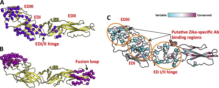
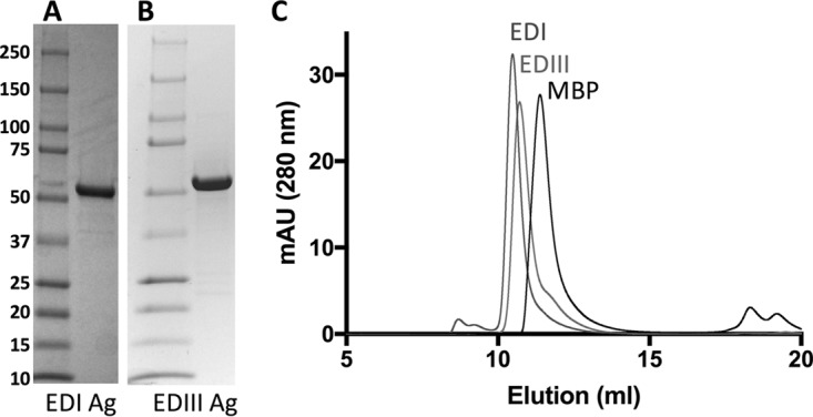
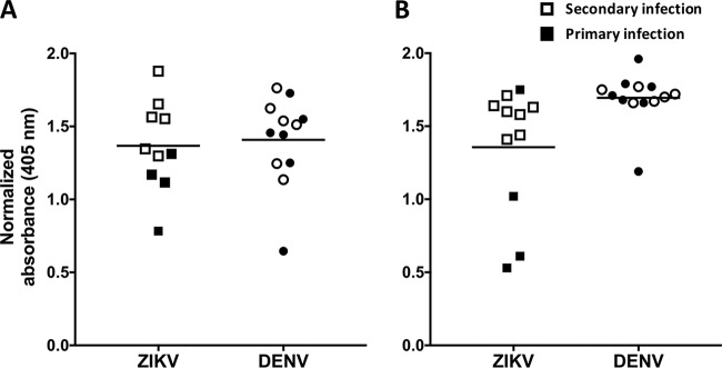
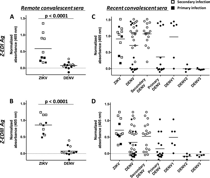
Zika virus (ZIKV) is an emerging flavivirus that can cause birth defects and neurologic complications. Molecular tests are effective for diagnosing acute ZIKV infection, although the majority of infections produce no symptoms at all or present after the narrow window in which molecular diagnostics are dependable. Serology is a reliable method for detecting infections after the viremic period; however, most serological assays have limited specificity due to cross-reactive antibodies elicited by flavivirus infections. Since ZIKV and dengue virus (DENV) widely cocirculate, distinguishing ZIKV infection from DENV infection is particularly important for diagnosing individual cases or for surveillance to coordinate public health responses. Flaviviruses also elicit type-specific antibodies directed to non-cross-reactive epitopes of the infecting virus; such epitopes are attractive targets for the design of antigens for development of serological tests with greater specificity. Guided by comparative epitope modeling of the ZIKV envelope protein, we designed two recombinant antigens displaying unique antigenic regions on domain I (Z-EDI) and domain III (Z-EDIII) of the ZIKV envelope protein. Both the Z-EDI and Z-EDIII antigens consistently detected ZIKV-specific IgG in ZIKV-immune sera but not cross-reactive IgG in DENV-immune sera in late convalescence (>12 weeks postinfection). In contrast, during early convalescence (2 to 12 weeks postinfection), secondary DENV-immune sera and some primary DENV-immune sera cross-reacted with the Z-EDI and Z-EDIII antigens. Analysis of sequential samples from DENV-immune individuals demonstrated that Z-EDIII cross-reactivity peaked in early convalescence and declined steeply over time. The Z-EDIII antigen has much potential as a diagnostic antigen for population-level surveillance and for detecting past infections in patients.

Keywords: ELISA; Zika virus; antibody-binding region; comparative epitope mapping; computational prediction; cross-reactivity; dengue virus; flavivirus; serological diagnosis; surveillance.


Figures

FIG 1
FIG 1
Identification of putative virus-specific antigenic regions on ZIKV E protein. We performed mapping of type-specific (A) and cross-reactive (B) epitopes on E protein by using experimentally determined antibody complex structures available in the Protein Data Bank. Contact residues observed at the interface between E protein and antibody in the complexes are shown as spheres (purple or magenta). (C) Mapping of the degrees of conservation of amino acid positions among eight clinically relevant flaviviruses. The color scale (cyan, variable region; and maroon, conserved region), as described in ConSurf (33, 51), is shown at the top. Three highly variable regions that overlap type-specific antibody-binding regions in panel A were identified as putative ZIKV-specific antibody-binding regions (orange circles), and the corresponding amino acid residues within this region are shown as spheres.
FIG 2
FIG 2
Analysis of purified recombinant antigens by SDS-PAGE and size exclusion chromatography (SEC). Purified Z-EDI (A) and Z-EDIII (B) antigens (6 μg/lane) were subjected to SDS-PAGE under reducing conditions and then stained with Coomassie brilliant blue. Molecular size markers and their apparent masses are shown on the left. (C) SEC overlays of purified EDI, EDIII, and MBP antigens. Protein samples in PBS were subjected to SEC on a Superdex75 10/300GL column. mAU, milli-absorbance units.
FIG 3
FIG 3
Binding of recombinant E80 antigen to sera from patients with remote (A) and recent (B) ZIKV and/or DENV infection. Sera from primary (filled symbols) and secondary (unfilled symbols) ZIKV- and DENV-infected patients were diluted 1:20, and the IgG antibodies bound to recombinant E80 antigen were measured by ELISA. Sera collected ≥12 weeks after infection were defined as remote infections, and sera collected within the first 12 weeks were considered to represent recent infections. The horizontal lines represent the means.
FIG 4
FIG 4
Binding of Z-EDI and Z-EDIII with remote (A and B) and recent (C and D) convalescent-phase sera from patients infected with ZIKV and/or DENV. Primary (filled symbols) and secondary (unfilled symbols) human serum samples were diluted 1:20, and the IgG antibodies bound to Z-EDI (A and C) or Z-EDIII (B and D) were measured using a sandwich ELISA. Sera collected ≥12 weeks after infection were defined as remote infections, and sera collected within the first 12 weeks were considered to represent recent infections. Statistical significances are indicated at the top of the graphs (Mann-Whitney U test). P values of <0.0001 were considered statistically significant. The horizontal lines represent the means.
FIG 5
FIG 5
Patterns of cross-reactivity of Z-EDIII (A) and ZED-I (B) antigens with longitudinal DENV samples. Intensities of serum binding in sandwich ELISAs for Z-EDIII and Z-EDI are shown as heat maps. Longitudinal samples (collected 14 to 23 days, 6 months, and 12 months postinfection) from patients with primary (left of the dashed line) or secondary (right of the dashed line) DENV3 infection were diluted 1:20, and the IgG antibodies bound to Z-EDI or Z-EDIII were measured using a sandwich ELISA. The resulting normalized OD values are represented by a color scale (green, lowest values; yellow, middle values; and red, highest values).

Similar articles

Cited by

References

    1. Musso D, Baud D, Gubler DJ. 2016. Zika virus: what do we know? Clin Microbiol Infect 22:494–496. doi:10.1016/j.cmi.2016.03.032. - DOI - PubMed
    1. Lazear HM, Diamond MS. 2016. Zika virus: new clinical syndromes and its emergence in the Western Hemisphere. J Virol 90:4864–4875. doi:10.1128/JVI.00252-16. - DOI - PMC - PubMed
    1. Lazear HM, Stringer EM, de Silva AM. 2016. The emerging Zika virus epidemic in the Americas: research priorities. JAMA 315:1945–1946. doi:10.1001/jama.2016.2899. - DOI - PubMed
    1. Cugola FR, Fernandes IR, Russo FB, Freitas BC, Dias JL, Guimaraes KP, Benazzato C, Almeida N, Pignatari GC, Romero S, Polonio CM, Cunha I, Freitas CL, Brandao WN, Rossato C, Andrade DG, Faria DP, Garcez AT, Buchpigel CA, Braconi CT, Mendes E, Sall AA, Zanotto PM, Peron JP, Muotri AR, Beltrao-Braga PC. 2016. The Brazilian Zika virus strain causes birth defects in experimental models. Nature 534:267–271. doi:10.1038/nature18296. - DOI - PMC - PubMed
    1. Garcez PP, Loiola EC, Madeiro da Costa R, Higa LM, Trindade P, Delvecchio R, Nascimento JM, Brindeiro R, Tanuri A, Rehen SK. 2016. Zika virus impairs growth in human neurospheres and brain organoids. Science 352:816–818. doi:10.1126/science.aaf6116. - DOI - PubMed

Publication types

MeSH terms