. 2018 Jan 31;38(5):1264-1276.
doi: 10.1523/JNEUROSCI.2869-17.2017. Epub 2017 Dec 20.

Selective Suppression of Local Interneuron Circuits in Human Motor Cortex Contributes to Movement Preparation

Affiliations

Selective Suppression of Local Interneuron Circuits in Human Motor Cortex Contributes to Movement Preparation

Ricci Hannah et al. J Neurosci. .

Abstract

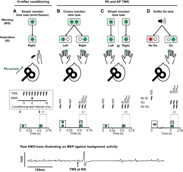
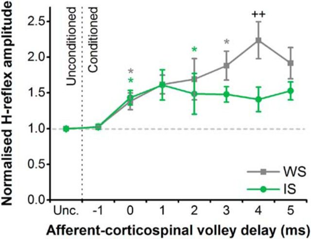
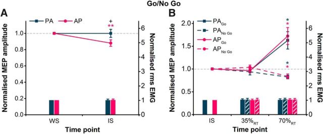
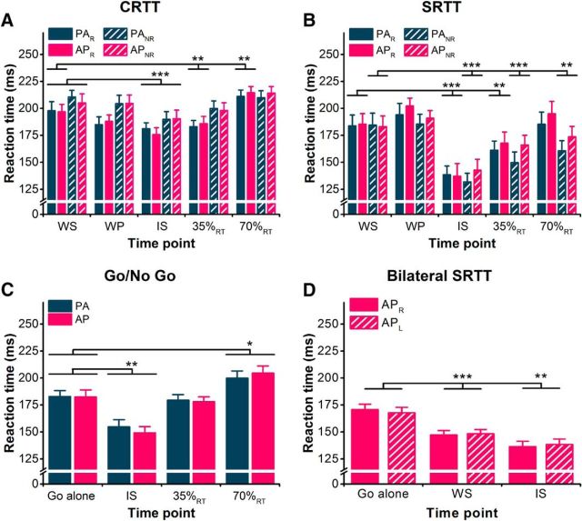
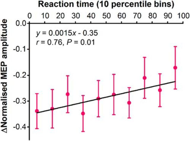
Changes in neural activity occur in the motor cortex before movement, but the nature and purpose of this preparatory activity is unclear. To investigate this in the human (male and female) brain noninvasively, we used transcranial magnetic stimulation (TMS) to probe the excitability of distinct sets of excitatory inputs to corticospinal neurons during the warning period of various reaction time tasks. Using two separate methods (H-reflex conditioning and directional effects of TMS), we show that a specific set of excitatory inputs to corticospinal neurons are suppressed during motor preparation, while another set of inputs remain unaffected. To probe the behavioral relevance of this suppression, we examined whether the strength of the selective preparatory inhibition in each trial was related to reaction time. Surprisingly, the greater the amount of selective preparatory inhibition, the faster the reaction time was. This suggests that the inhibition of inputs to corticospinal neurons is not involved in preventing the release of movement but may in fact facilitate rapid reactions. Thus, selective suppression of a specific set of motor cortical neurons may be a key aspect of successful movement preparation.SIGNIFICANCE STATEMENT Movement preparation evokes substantial activity in the motor cortex despite no apparent movement. One explanation for the lack of movement is that motor cortical output in this period is gated by an inhibitory mechanism. This notion was supported by previous noninvasive TMS studies of human motor cortex indicating a reduction of corticospinal excitability. On the contrary, our data support the idea that there is a coordinated balance of activity upstream of the corticospinal output neurons. This includes a suppression of specific local circuits that supports, rather than inhibits, the rapid generation of prepared movements. Thus, the selective suppression of local circuits appears to be an essential part of successful movement preparation instead of an external control mechanism.

Keywords: corticospinal; inhibition; motor cortex; motor preparation; transcranial magnetic stimulation.


Figures

Figure 1.
Figure 1.
Reaction time tasks and stimulus timings. A, For the SRTT in experiment 1, participants performed the task with their right wrist, and median nerve stimulus (MNS) and TMS stimulus timings were limited to WS and IS time points. B, For the CRTT in experiment 2, a noninformative visual WS (left and right LEDs lit for 150 ms) preceded a left or right IS (75 ms duration), which cued a response with either the left or right index, respectively. C, In experiment 3, participants performed separate blocks of the SRTT with their left and right index fingers. They received a visual WS (150 ms duration) before a visual IS (75 ms duration). D, For the Go/No Go task in experiment 4, an auditory WS (500 Hz tone, 150 ms duration) preceded either a green (Go) or red (No Go) visual stimulus (75 ms duration), which cued the execution of a right index response and the withholding of a response, respectively. Within each experiment, stimuli were delivered at one of several time points in a trial: at the WS, in the warning period (WP) 0.25 s after the WS and before the IS (B, C) at the IS and after the IS at 35% and 70% of the mean baseline reaction time (35%RT, 70%RT). TMS was delivered with the coil positioned to induce PA currents (see A) only in experiment 1, and both PA and AP currents (position coil handle rotated 180° around the intersection of coil windings) in experiments 2–4. Note that for trials cueing a right-hand response, MEPs were recorded from the (right) responding hand; and for trials cueing a left-hand response, MEPs were recorded from the (right) nonresponding hand. An example raw EMG trace is shown at the bottom to illustrate the MEP against the background voluntary muscle activity during experiments 2–5.
Figure 2.
Figure 2.
H-reflexes conditioned with TMS during the simple reaction time task. The interval between the conditioning TMS stimulus and the test H-reflex stimulus that produced coincident arrival of the corticospinal and afferent volleys at the spinal motoneurons, and thus facilitated the H-reflex, was considered to be 0 ms (i.e., the afferent–corticospinal volley delay is 0). Positive values for the delay (e.g., +1 ms) then reflected delayed arrival of the afferent compared with corticospinal volleys, while negative values (e.g., −1 ms) reflected the earlier arrival of the afferent volleys compared with the corticospinal volleys. During the simple reaction time task, H-reflexes in the FCR muscle were facilitated to a lesser extent at the IS than the WS, specifically when the arrival of the afferent volleys at the spinal motoneurons was delayed relative to the corticospinal volleys (4 ms). By contrast, H-reflexes were facilitated to a similar extent at the IS and WS when the afferent and corticospinal volleys arrived coincidentally at the spinal motoneurons (0 ms). *p < 0.05, compared with unconditioned (Unc.) H-reflex within each time point (WS and IS); ++p < 0.01, IS vs WS.
Figure 3.
Figure 3.
A, B, During the choice reaction time task, MEP amplitudes in the right FDI shown normalized to the WS time point (colored lines, left y-axis) were suppressed more for AP currents than for PA currents at the IS during right hand-responding trials (A) and at the IS and 70%RT in right-hand nonresponding trials (B). The facilitation of MEPs in right hand-responding trials at 70%RT was similar for both current directions (A). Voluntary rms EMG (colored bars, right y-axis) measured before the TMS pulses is shown normalized to values at the WS, and was similar for PA and AP currents across different time points for right hand-responding (A) and nonresponding trials (B). C, D, MEP latencies were longer for AP currents compared with PA currents in both right hand-responding (C) and nonresponding (D) trials at all time points except 70%RT in responding trials. **p < 0.01, ***p < 0.001, compared with the WS time point within each current direction; ++p < 0.01, +++p < 0.001, AP vs PA.
Figure 4.
Figure 4.
A, During the simple reaction time task, MEP amplitudes in the right FDI shown normalized to the WS time point (colored lines, left y-axis) and were suppressed more for AP currents than for PA currents at the IS and 35%RT during right hand-responding blocks. The facilitation of MEPs in the same block at 70%RT was similar for both current directions. B, However, for right-hand nonresponding blocks, normalized MEP amplitudes were suppressed to a similar extent for AP and PA currents at all times following the WS. Voluntary rms EMG (colored bars, right y-axis) measured before the TMS pulse is shown normalized to values at the WS, and was similar for PA and AP currents across different time points for right hand-responding (A) and nonresponding (B) blocks. C, MEP latencies measured at the WS were longer for AP currents compared with PA currents in both right hand-responding and nonresponding blocks. *p < 0.05, **p < 0.01, ***p < 0.001, compared with WS time point within each current direction; +p < 0.05, ++p < 0.01, +++p < 0.001, AP vs PA.
Figure 5.
Figure 5.
A, B, During the Go/No Go task, MEP amplitudes in the right FDI, shown normalized to the WS time point (colored lines, left y-axis), were suppressed more for AP currents than for PA currents at the IS compared with the WS (A), indicating a selective anticipatory suppression in response to the WS. However, during successful No Go trials of the Go/No Go task, MEP amplitudes normalized to the IS were suppressed to a similar extent for AP currents as for PA currents at 70%RT when compared with those at the IS (B), indicating a similar reactive suppression in response to the No Go signal. The facilitation of MEPs in Go trials at 70%RT was similar for both current directions. Voluntary rms EMG measured before the TMS pulse (colored bars, right y-axis) is shown normalized to values at the WS (A) and IS (B), and was similar for PA and AP currents across different time points for Go and No Go trials. *p < 0.05, **p < 0.01, compared with IS time point within each current direction; +p < 0.05, AP vs PA.
Figure 6.
Figure 6.
A–D, Mean EMG-determined reaction times shown for correct response trials and both PA and AP current directions in CRTT (A), SRTT (B), Go/No Go (C), and bilateral SRTT tasks (D). For the legends in A and B, subscript R denotes right hand-responding trials and subscript NR denotes right-hand nonresponding trials (i.e., reaction times determined from the left hand). For the legend in D, subscript R and L denote right-hand and left-hand responses in the same trial. *p < 0.05, **p < 0.01, ***p < 0.001, compared with WS time point in A and B and to Go alone (C, D).
Figure 7.
Figure 7.
Correlation between mean MEP amplitude change and simple reaction time arranged in consecutive 10 percentile bins (e.g., 0 to 10th, 10th to 20th).

Comment in

Similar articles

Cited by

References

    1. Badry R, Mima T, Aso T, Nakatsuka M, Abe M, Fathi D, Foly N, Nagiub H, Nagamine T, Fukuyama H (2009) Suppression of human cortico-motoneuronal excitability during the Stop-signal task. Clin Neurophysiol 120:1717–1723. 10.1016/j.clinph.2009.06.027 - DOI - PubMed
    1. Bestmann S, Duque J (2016) Transcranial magnetic stimulation: decomposing the process underlying action preparation. Neuroscientist 22:392–405. 10.1177/1073858415592594 - DOI - PubMed
    1. Churchland MM, Cunningham JP, Kaufman MT, Ryu SI, Shenoy KV (2010) Cortical preparatory activity: representation of movement or first cog in a dynamical machine? Neuron 68:387–400. 10.1016/j.neuron.2010.09.015 - DOI - PMC - PubMed
    1. Day BL, Dressler D, Maertens de Noordhout A, Marsden CD, Nakashima K, Rothwell JC, Thompson PD (1989a) Electric and magnetic stimulation of human motor cortex: surface EMG and single motor unit responses. J Physiol 412:449–473. 10.1113/jphysiol.1989.sp017626 - DOI - PMC - PubMed
    1. Day BL, Rothwell JC, Thompson PD, Maertens De Noordhout A, Nakashima K, Shannon K, Marsden CD (1989b) Delay in the execution of voluntary movement by electrical or magnetic brain stimulation in intact man: evidence for the storage of motor programs in the brain. Brain 112:649–663. 10.1093/brain/112.3.649 - DOI - PubMed

Publication types

LinkOut - more resources