. 2018 Jan 31;38(5):1151-1159.
doi: 10.1523/JNEUROSCI.1975-17.2017. Epub 2017 Dec 20.

Cocaine Selectively Reorganizes Excitatory Inputs to Substantia Nigra Pars Compacta Dopamine Neurons

Affiliations

Cocaine Selectively Reorganizes Excitatory Inputs to Substantia Nigra Pars Compacta Dopamine Neurons

Gerard M J Beaudoin 3rd et al. J Neurosci. .

Abstract

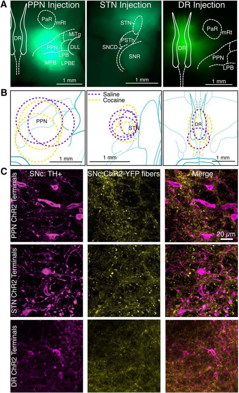
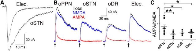
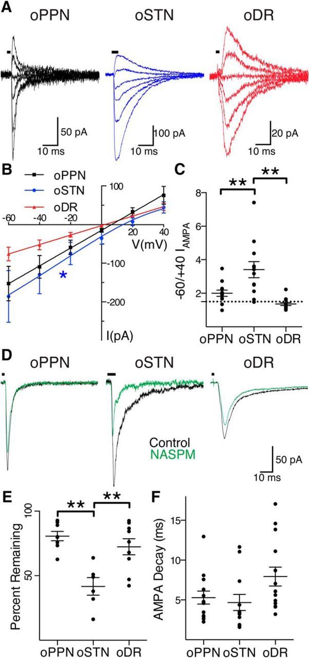
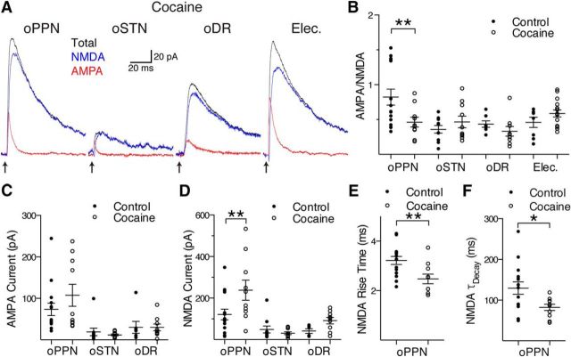
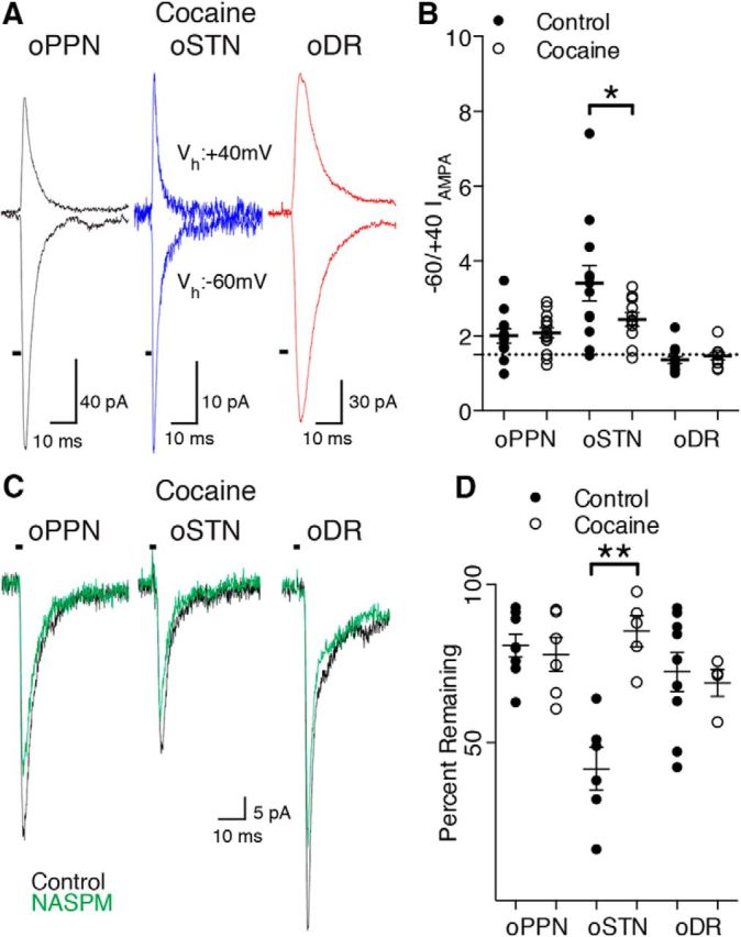
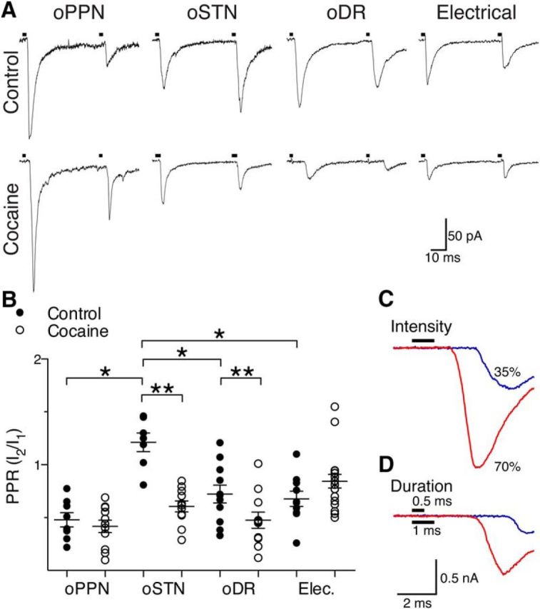
Substantia nigra pars compacta (SNc) dopamine neurons and their targets are involved in addiction and cue-induced relapse. However, afferents onto SNc dopamine neurons themselves appear insensitive to drugs of abuse, such as cocaine, when afferents are collectively stimulated electrically. This contrasts with ventral tegmental area (VTA) dopamine neurons, whose glutamate afferents react robustly to cocaine. We used an optogenetic strategy to isolate identified SNc inputs and determine whether cocaine sensitivity in the mouse SNc circuit is conferred at the level of three glutamate afferents: dorsal raphé nucleus (DR), pedunculopontine nucleus (PPN), and subthalamic nucleus (STN). We found that excitatory afferents to SNc dopamine neurons are sensitive to cocaine in an afferent-specific manner. A single exposure to cocaine in vivo led to PPN-innervated synapses reducing the AMPA-to-NMDA receptor-mediated current ratio. In contrast to work in the VTA, this was due to increased NMDA receptor function with no change in AMPA receptor function. STN synapses showed a decrease in calcium-permeable AMPA receptors after cocaine, but no change in the AMPA-to-NMDA ratio. Cocaine also increased the release probability at DR-innervated and STN-innervated synapses, quantified by decreases in paired-pulse ratios. However, release probability at PPN-innervated synapses remained unaffected. By examining identified inputs, our results demonstrate a functional distribution among excitatory SNc afferent nuclei in response to cocaine, and suggest a compelling architecture for differentiation and separate parsing of inputs within the nigrostriatal system.SIGNIFICANCE STATEMENT Prior studies have established that substantia nigra pars compacta (SNc) dopamine neurons are a key node in the circuitry that drives addiction and relapse, yet cocaine apparently has no effect on electrically stimulated excitatory inputs. Our study is the first to demonstrate the functional impact of a drug of abuse on synaptic mechanisms of identified afferents to the SNc. Optogenetic dissection of inputs originating from dorsal raphé, pedunculopontine, and subthalamic nuclei were tested for synaptic modifications following in vivo cocaine exposure. Our results demonstrate that cocaine differentially induces modifications to SNc synapses depending on input origin. This presents implications for understanding dopamine processing of motivated behavior; most critically, it indicates that dopamine neurons selectively modulate signal reception processed by afferent nuclei.

Keywords: AMPA; NMDA; dorsal raphé; optogenetics; pedunculopontine nucleus; subthalamic nucleus.


Figures

Figure 1.
Figure 1.
Injection of adeno-associated virus-encoded ChR2-YFP in the DR, PPN, and STN targets YFP expression to each nucleus and its projection targets. A, Horizontal sections from mice injected with virus into PPN, STN, and DR, demonstrate YFP expression (green) throughout the nucleus 4 weeks after injection. DLL, Dorsal nucleus of the lateral meniscus; LPB, lateral parabrachial nucleus; LPBE, LPB external part; MiTg, microcellular tegmental nucleus; MPB, medial parabrachial nucleus; mRt, mesencephalic reticular formation; PaR, pararubral nucleus; PSTh, parasubthalamic nucleus; SNCD, substantia nigra, compact part, dorsal tier; SNR, substantia nigra, reticular part. B, Ovals represent the distribution of labeled neurons for mice used for control (magenta) and cocaine-injected (yellow) mice. C, YFP-labeled afferents (yellow) from PPN, STN, and DR are seen among anti-tyrosine hydroxylase-labeled dopamine neurons (magenta) in SNC.
Figure 2.
Figure 2.
PPN, STN, and DR afferents have distinct postsynaptic receptor compositions in drug-naive mice. A, Example of a matched optogenetically evoked EPSC in STN with an electrically evoked EPSC in the same dopamine neuron. B, Optogenetically evoked EPSCs (black traces; stimulation for all traces at arrows) from PPN (oPPN), STN (oSTN), DR (oDR), and electrically evoked EPSCs (Elec.; stimulus artifact removed) contain both NMDA (blue traces) and AMPA (red traces) receptor-mediated currents. C, AMPA-to-NMDA receptor-mediated current ratios. oPPN-evoked EPSCs had similar amount of AMPA receptor-mediated and NMDA receptor-mediated currents with a ratio near 1. oSTN-evoked, oDR-evoked, and electrically evoked EPSCs have AMPA-to-NMDA receptor-mediated current ratios of <0.5. *p < 0.05, **p < 0.01.
Figure 3.
Figure 3.
PPN, STN, and DR afferents activate different complements of AMPA receptors on dopamine neurons. A, oPPN-evoked (black), oSTN-evoked (blue), and oDR-evoked (red) AMPA receptor-mediated currents at holding voltages of −60, −40, −20, 0, +20, and +40 mV, from bottom trace to top trace. B, oPPN-evoked and oDR-evoked AMPA receptor-mediated currents (black and red) have a linear I–V relationship, indicating that they contain the GluA2 subunit. oSTN-evoked AMPA receptor-mediated current with inwardly rectifying I–V relationship (blue), not going through 0 pA, 0 mV. C, Rectification index corresponding to the ratio of currents at a holding voltage of −60 to +40 mV. The dashed line represents a ratio of 1.5, corresponding to a linear I–V relationship. D, oPPN evoked an inward current (black) that is marginally sensitive to NASPM (green). oSTN-evoked AMPA receptor-mediated current (black) is significantly inhibited by NASPM (green), indicating the presence of calcium-permeable AMPA receptors. oDR-evoked AMPA receptor-mediated current (black) is also partially inhibited by NASPM (green). E, Calcium-permeable AMPA receptor antagonists JTx and NASPM significantly decrease the normalized amplitude of AMPA receptor-mediated currents from oSTN compared with oPPN and oDR. F, The time constant of decay for AMPA receptor-mediated currents produced at a holding voltage of −60 mV. *p < 0.05, **p < 0.01.
Figure 4.
Figure 4.
Exposure to cocaine decreases the proportion of AMPA receptors relative to NMDA receptors at PPN synapses by changing the NMDA receptor content. A, After a single exposure to cocaine in vivo, oPPN-evoked AMPA (red) receptor-mediated and NMDA (blue) receptor-mediated currents become similar to oSTN-evoked, oDR-evoked, and electrically evoked (Elec.; stimulus artifact removed) currents. The oPPN-evoked EPSC and electrically evoked EPSC were recorded from the same dopamine neuron. Total EPSC is indicated by the traces in black. B, Cocaine decreases the ratio of AMPA-to-NMDA currents at PPN inputs, but does not affect the AMPA-to-NMDA ratio of oSTN-evoked, oDR-evoked, or electrically evoked EPSCs. C, The amplitude of the AMPA receptor-mediated EPSCs evoked from oPPN, oSTN, and oDR, are not significantly affected after exposure to cocaine. D, Exposure to cocaine significantly increases the amplitude of NMDA receptor-mediated currents evoked by oPPN, but not oSTN or oDR. E, F, NMDA receptor-mediated EPSCs evoked by oPPN show a reduced rise time (E) and time constant of decay (F) after cocaine administration. *p < 0.05, **p < 0.01.
Figure 5.
Figure 5.
A single injection of cocaine in vivo causes removal of calcium-permeable AMPA receptors at STN synapses. A, After exposure to cocaine, oPPN-evoked (black), oSTN-evoked (blue), and oDR-evoked (red) EPSCs have similar rectification of AMPA receptor-mediated currents. B, Rectification index of AMPA receptor-mediated currents from individual afferents in control (saline and naive) and cocaine-treated animals. The dashed line represents a ratio of 1.5, corresponding to a linear I–V relationship. C, After exposure to cocaine, oPPN-evoked and oDR-evoked inward currents (black) are partially sensitive to NASPM (green). After cocaine injection, oSTN-evoked AMPA receptor-mediated current (black) is only partially sensitive to NASPM (green), indicating the relative decrease of calcium-permeable AMPA receptors when compared with the same input in control animals. D, Calcium-permeable AMPA receptor antagonists JTx and NASPM do not significantly decrease the normalized amplitude of AMPA receptor-mediated currents from the oSTN, oDR, or oPPN after cocaine exposure in vivo. *p < 0.05, **p < 0.01.
Figure 6.
Figure 6.
Cocaine increases the probability of release at STN and DR synapses but does not affect PPN synapses. A, oPPN-evoked and oDR-evoked EPSCs demonstrate paired-pulse depression. oSTN-evoked EPSCs show paired-pulse facilitation whereas electrical stimulation (artifact-blanked) evoked EPSCs with paired-pulse depression. Exposure to cocaine leads to paired-pulse depression from facilitation of oSTN-evoked EPSCs, increased paired-pulse depression of oDR-evoked EPSCs, and no change in oPPN-evoked or electrically evoked EPSCs. B, oSTN-evoked EPSCs have a PPR that is significantly higher than electrically evoked or oPPN-evoked EPSCs. The PPR of oDR-evoked and oSTN-evoked EPSCs is significantly reduced by in vivo cocaine exposure, while PPRs of electrically evoked and oPPN-evoked EPSCs remain unchanged. C, D, An example of oPPN-evoked EPSCs in which amplitude increased and onset latency decreased as the light intensity (C) or pulse duration (D) was increased, indicating the EPSCs were elicited by propagating action potentials. *p < 0.05, **p < 0.01.

Similar articles

Cited by

References

    1. Argilli E, Sibley DR, Malenka RC, England PM, Bonci A (2008) Mechanism and time course of cocaine-induced long-term potentiation in the ventral tegmental area. J Neurosci 28:9092–9100. 10.1523/JNEUROSCI.1001-08.2008 - DOI - PMC - PubMed
    1. Baunez C, Dias C, Cador M, Amalric M (2005) The subthalamic nucleus exerts opposite control on cocaine and ‘natural’ rewards. Nat Neurosci 8:484–489. 10.1038/nn1429 - DOI - PubMed
    1. Bechara A, van der Kooy D (1989) The tegmental pedunculopontine nucleus: a brain-stem output of the limbic system critical for the conditioned place preferences produced by morphine and amphetamine. J Neurosci 9:3400–3409. - PMC - PubMed
    1. Bellone C, Lüscher C (2006) Cocaine triggered AMPA receptor redistribution is reversed in vivo by mGluR-dependent long-term depression. Nat Neurosci 9:636–641. 10.1038/nn1682 - DOI - PubMed
    1. Bowie D, Mayer ML (1995) Inward rectification of both AMPA and kainate subtype glutamate receptors generated by polyamine-mediated ion channel block. Neuron 15:453–462. 10.1016/0896-6273(95)90049-7 - DOI - PubMed

Publication types

MeSH terms

LinkOut - more resources