. 2018 Mar;364(3):494-503.
doi: 10.1124/jpet.117.244129. Epub 2017 Dec 20.

Triple Angiokinase Inhibitor Nintedanib Directly Inhibits Tumor Cell Growth and Induces Tumor Shrinkage via Blocking Oncogenic Receptor Tyrosine Kinases

Affiliations

Triple Angiokinase Inhibitor Nintedanib Directly Inhibits Tumor Cell Growth and Induces Tumor Shrinkage via Blocking Oncogenic Receptor Tyrosine Kinases

Frank Hilberg et al. J Pharmacol Exp Ther. 2018 Mar.

Abstract

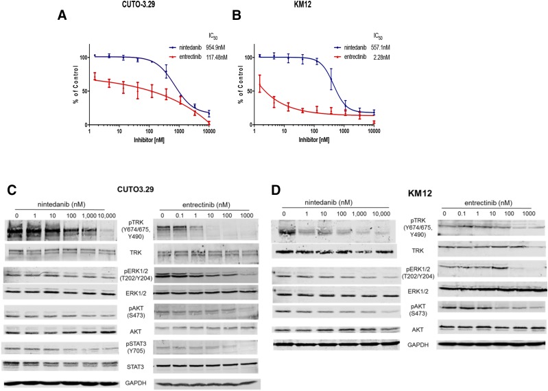
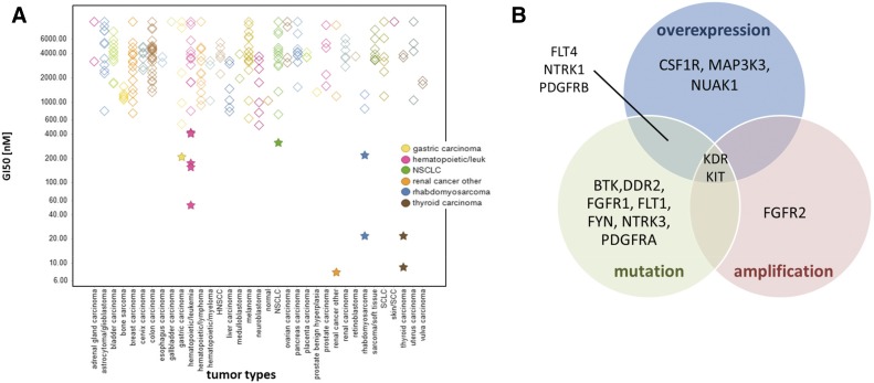
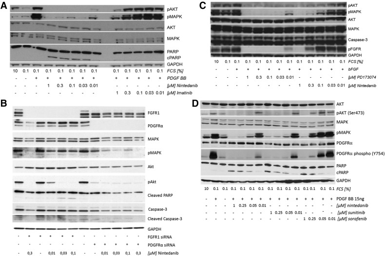
The triple-angiokinase inhibitor nintedanib is an orally available, potent, and selective inhibitor of tumor angiogenesis by blocking the tyrosine kinase activities of vascular endothelial growth factor receptor (VEGFR) 1-3, platelet-derived growth factor receptor (PDGFR)-α and -β, and fibroblast growth factor receptor (FGFR) 1-3. Nintedanib has received regulatory approval as second-line treatment of adenocarcinoma non-small cell lung cancer (NSCLC), in combination with docetaxel. In addition, nintedanib has been approved for the treatment of idiopathic lung fibrosis. Here we report the results from a broad kinase screen that identified additional kinases as targets for nintedanib in the low nanomolar range. Several of these kinases are known to be mutated or overexpressed and are involved in tumor development (discoidin domain receptor family, member 1 and 2, tropomyosin receptor kinase A (TRKA) and C, rearranged during transfection proto-oncogene [RET proto oncogene]), as well as in fibrotic diseases (e.g., DDRs). In tumor cell lines displaying molecular alterations in potential nintedanib targets, the inhibitor demonstrates direct antiproliferative effects: in the NSCLC cell line NCI-H1703 carrying a PDGFRα amplification (ampl.); the gastric cancer cell line KatoIII and the breast cancer cell line MFM223, both driven by a FGFR2 amplification; AN3CA (endometrial carcinoma) bearing a mutated FGFR2; the acute myeloid leukemia cell lines MOLM-13 and MV-4-11-B with FLT3 mutations; and the NSCLC adenocarcinoma LC-2/ad harboring a CCDC6-RET fusion. Potent kinase inhibition does not, however, strictly translate into antiproliferative activity, as demonstrated in the TRKA-dependent cell lines CUTO-3 and KM-12. Importantly, nintedanib treatment of NCI-H1703 tumor xenografts triggered effective tumor shrinkage, indicating a direct effect on the tumor cells in addition to the antiangiogenic effect on the tumor stroma. These findings will be instructive in guiding future genome-based clinical trials of nintedanib.


Figures

Fig. 1.
Fig. 1.
Inhibition of proliferation and TRKA and downstream signaling of TRKA fusion cell lines using nintedanib and entrectinib. Human TRKA fusion cell lines CUTO3.29 (A) and KM12 (B) were treated with the indicated dose range of ninitedanib (blue) or entrectinib (red) and assayed for cell proliferation 72 hours after treatment using an MTS assay (n = 3, error bars represent ± S.E.M.). The IC50 was calculated for each cell line as follows: CUTO3.29 treated with nintedanib, 954.9 ± 7.2 nmol/l; or with entrectinib, 117.48 ± 5.6 nmol/liter; KM12 treated with nintedanib, 557.1 ± 5.6 nmol/l; and with entrectinib, 2.28 ± 0.3 nmol/l. CUTO3.29 (C) and KM12 (D) were treated with the indicated increasing doses of either nintedanib or entrectinib, and protein lysates were collected 2 hours later for Western blot analysis of phospho-TRKA and downstream signaling of phospho-ERK1/2, phospho-AKT and phospho-STAT3. Western blot images are representative of three independent experiments.
Fig. 2.
Fig. 2.
Mutational analysis of nintedanib sensitive tumor cell lines. (A) Response of Ricerca 240-OncoPanel cell lines to nintedanib. *Sensitive cell lines (GI50 ≤ 500 nmol/l). The color code legend highlights the tumor types represented by the sensitive cell lines. (B) Venn diagram showing the 16 genes and their respective alterations in the sensitive cell lines highlighted in (A).
Fig. 3.
Fig. 3.
Nintedanib inhibits ligand-dependent phosphorylation of MAPK and Akt in NCI-H1703 and KatoIII tumor cells. (A) NCI-H1703 Western blot analysis after exposure to either nintedanib or imatinib after stimulation with PDGF BB. Strong concentration-dependent reduction of phosphorylated MAPK and Akt levels by nintedanib compared with imatinib. (B) NCI-H1703 Western blot analysis after 72 hours of either FGFR1 or PDGFRA siRNA treatment and 2 hours of exposure to nintedanib shows concentration-dependent reduction of phosphorylated MAPK and Akt levels. Cleaved PARP and cleaved caspase-3 serve as indicators for apoptosis. Nintedanib concentrations are shown in micromoles per liter. (C) KatoIII Western blot analysis after exposure to either nintedanib or after stimulation with bFGF. Slightly stronger concentration-dependent reduction of phosphorylated MAPK and Akt levels by PD173074 compared with nintedanib. Western blot images are representative of three independent experiments. (D) NCI-H1703 Western blot analysis after exposure to nintedanib, sunitinib, or sorafenib after stimulation with PDGF BB. Concentration-dependent reduction of phosphorylated MAPK, Akt, and phosphor PDGFRα levels by nintedanib and sunitinib compared with sorafenib.
Fig. 4.
Fig. 4.
Single-agent nintedanib induces tumor shrinkage of subcutaneous NCI-H1703 tumors. (A) Median tumor volume over time, (B) Single tumor volumes + median at day 29; nintedanib, 100 mg/kg daily (open triangles); vatalanib, 100 mg/kg daily (stars); imatinib, 75 mg/kg daily (filled triangles); and imatinib, 75 mg/kg twice daily (filled diamonds) (n = 7). Nintedanib in combination with the BET inhibitor BI894999 shows excellent antitumor efficacy, with seven of eight animals surviving ≥100 days. (C) Median tumor volume over time. (Nintedanib-treated animals euthanized prematurely because of tumor necrosis.) (D) Single tumor volumes + median at day 33. (E) Survival probability (n = 8 for treatment, n = 10 for vehicle control). Xenografts were established subcutaneously in athymic mice and allowed to reach a volume of ∼100 mm3 before treatment.

References

    1. Alioto TS, Buchhalter I, Derdak S, Hutter B, Eldridge MD, Hovig E, Heisler LE, Beck TA, Simpson JT, Tonon L, et al. (2015) A comprehensive assessment of somatic mutation detection in cancer using whole-genome sequencing. Nat Commun 6:10001. - PMC - PubMed
    1. Bengtsson H, Irizarry R, Carvalho B, Speed TP. (2008) Estimation and assessment of raw copy numbers at the single locus level. Bioinformatics 24:759–767. - PubMed
    1. Bengtsson H, Wirapati P, Speed TP. (2009) A single-array preprocessing method for estimating full-resolution raw copy numbers from all Affymetrix genotyping arrays including GenomeWideSNP 5 & 6. Bioinformatics 25:2149–2156. - PMC - PubMed
    1. Berge EM, Doebele RC. (2014) Targeted therapies in non-small cell lung cancer: emerging oncogene targets following the success of epidermal growth factor receptor. Semin Oncol 41:110–125. - PMC - PubMed
    1. Blackhall F, Cappuzzo F. (2016) Crizotinib: from discovery to accelerated development to front-line treatment. Ann Oncol 27 (Suppl 3):iii35–iii41. - PubMed

Publication types

LinkOut - more resources