. 2018 Feb 26;92(6):e01535-17.
doi: 10.1128/JVI.01535-17. Print 2018 Mar 15.

Identification of Residues Controlling Restriction versus Enhancing Activities of IFITM Proteins on Entry of Human Coronaviruses

Affiliations

Identification of Residues Controlling Restriction versus Enhancing Activities of IFITM Proteins on Entry of Human Coronaviruses

Xuesen Zhao et al. J Virol. .

Abstract

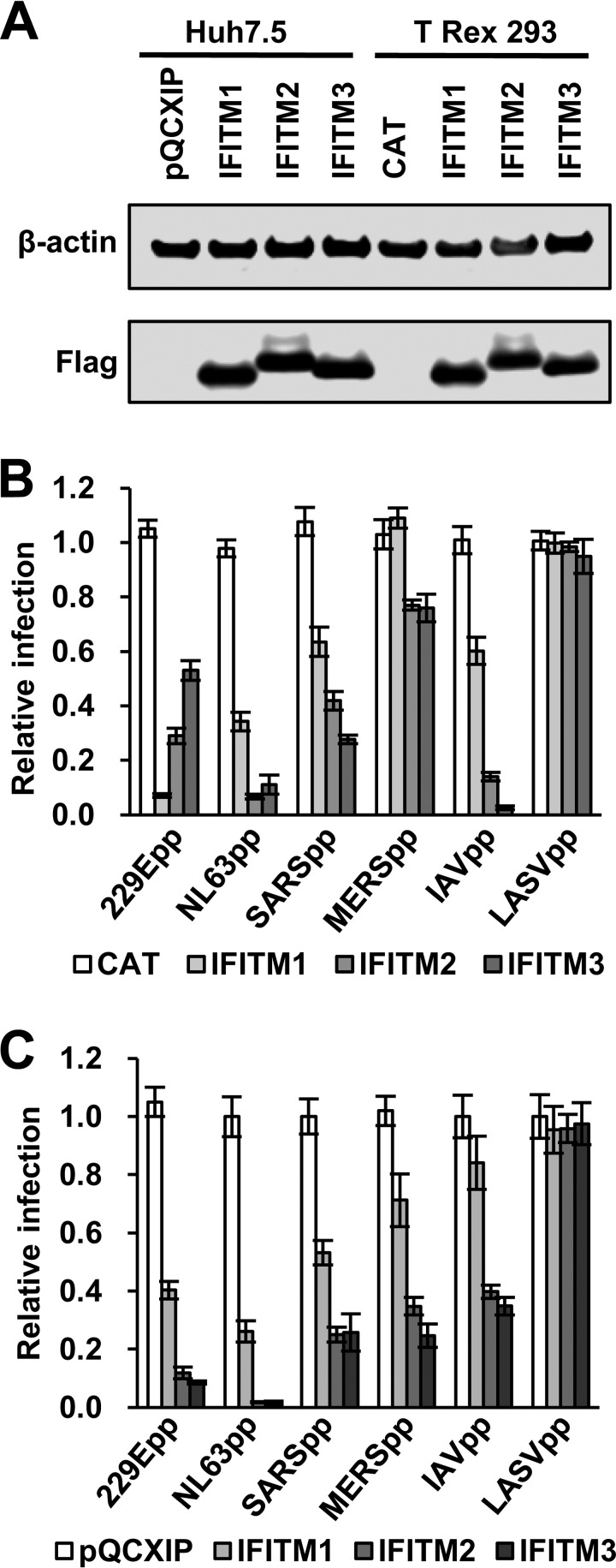
Interferon-induced transmembrane proteins (IFITMs) are restriction factors that inhibit the infectious entry of many enveloped RNA viruses. However, we demonstrated previously that human IFITM2 and IFITM3 are essential host factors facilitating the entry of human coronavirus (HCoV) OC43. In a continuing effort to decipher the molecular mechanism underlying IFITM differential modulation of HCoV entry, we investigated the roles of structural motifs important for IFITM protein posttranslational modifications, intracellular trafficking, and oligomerization in modulating the entry of five HCoVs. We found that three distinct mutations in IFITM1 or IFITM3 converted the host restriction factors to enhance entry driven by the spike proteins of severe acute respiratory syndrome coronavirus (SARS-CoV) and/or Middle East respiratory syndrome coronavirus (MERS-CoV). First, replacement of IFITM3 tyrosine 20 with either alanine or aspartic acid to mimic unphosphorylated or phosphorylated IFITM3 reduced its activity to inhibit the entry of HCoV-NL63 and -229E but enhanced the entry of SARS-CoV and MERS-CoV. Second, replacement of IFITM3 tyrosine 99 with either alanine or aspartic acid reduced its activity to inhibit the entry of HCoV-NL63 and SARS-CoV but promoted the entry of MERS-CoV. Third, deletion of the carboxyl-terminal 12 amino acid residues from IFITM1 enhanced the entry of MERS-CoV and HCoV-OC43. These findings suggest that these residues and structural motifs of IFITM proteins are key determinants for modulating the entry of HCoVs, most likely through interaction with viral and/or host cellular components at the site of viral entry to modulate the fusion of viral envelope and cellular membranes.IMPORTANCE The differential effects of IFITM proteins on the entry of HCoVs that utilize divergent entry pathways and membrane fusion mechanisms even when using the same receptor make the HCoVs a valuable system for comparative investigation of the molecular mechanisms underlying IFITM restriction or promotion of virus entry into host cells. Identification of three distinct mutations that converted IFITM1 or IFITM3 from inhibitors to enhancers of MERS-CoV or SARS-CoV spike protein-mediated entry revealed key structural motifs or residues determining the biological activities of IFITM proteins. These findings have thus paved the way for further identification of viral and host factors that interact with those structural motifs of IFITM proteins to differentially modulate the infectious entry of HCoVs.

Keywords: IFITM; coronavirus; viral entry.


Figures

FIG 1
FIG 1
IFITM proteins inhibit infection by pseudoviruses in FLIP-IN T Rex 293 and Huh7.5 cells. (A) The expression of FLAG-tagged IFITMs in FLIP-IN T Rex 293 cells cultured in the presence of 1 μg/ml of Tet for 24 h and in Huh7.5-derived stable cell lines was detected by a Western blot assay using a monoclonal antibody against FLAG tag. β-Actin served as a loading control. (B) FLIP-IN T Rex 293 cells expressing CAT or the indicated IFITM protein were cultured in the presence or absence of Tet for 24 h and then infected with 229Epp, NL63pp, SARSpp, MERSpp, IAVpp, or LASVpp. Luciferase activities were determined at 48 h postinfection (hpi). Relative infection efficiency is the ratio of luciferase activity in cells cultured in the presence of Tet over that in cells cultured in the absence of Tet. The error bars indicate standard deviations (n = 6). IFITMs did not significantly affect infection by LASVpp but significantly (P < 0.001) inhibited infection by all the other tested pseudotyped viruses. (C) Huh7.5 cells expressing N-terminally FLAG-tagged human IFITM proteins or transduced with an empty vector (pQCXIP) were infected with the indicated pseudoviruses. Luciferase activities were determined at 48 hpi. Relative infection is the ratio of the luciferase activity of cells expressing the indicated IFITM protein over that of cells transduced with the empty vector. The error bars indicate standard deviations (n = 6). All three IFITM proteins significantly (P < 0.001) inhibited infection by all the pseudoviruses except LASVpp.
FIG 2
FIG 2
Y20 of IFITM3 plays a critical role in modulating the entry of 229E, MERS-CoV, SARS-CoV, and NL63 in both 293 and Huh7.5 cells. (A) FLIP-IN T Rex cells expressing CAT or the indicated wild-type or mutant IFITM proteins were cultured in the presence of 1 μg/ml of Tet for 24 h. Expression of FLAG-tagged IFITM mutants was detected by Western blotting assay using anti-FLAG monoclonal antibody. β-Actin served as a loading control. (B to E) The above-mentioned FLP-IN T Rex-derived cell lines were cultured in the presence or absence of Tet for 24 h and then infected with HCoV-OC43pp (B), SARSpp or NL63pp (C), 229Epp (D), or MERSpp and LASVpp (E). Luciferase activities were determined at 48 hpi. Relative infection efficiency is the ratio of the luciferase activity in cells cultured in the presence of Tet over that in cells cultured in the absence of Tet. The error bars indicate standard deviations (n = 6). (F) Huh7.5 cells were stably transduced with an empty retroviral vector (pQCXIP) or vectors expressing wild-type or mutant IFITM3 proteins. Expression of FLAG-tagged IFITM proteins was detected by Western blotting assay using an anti-FLAG monoclonal antibody. β-Actin served as a loading control. (G to K) The above-mentioned Huh7.5-derived cell lines were infected with OC43pp (G), SARSpp and NL63pp (H), 229Epp (I), MERSpp (J), or LASVpp (K). Luciferase activities were determined at 48 hpi. Relative infection is the ratio of the luciferase activity of cells expressing the indicated IFITM protein over that of cells transduced with the empty vector. The error bars indicate standard deviations (n = 6).
FIG 3
FIG 3
Y20 mutation alters IFITM3 subcellular localization. FLP-IN T Rex cells expressing the indicated wild-type and mutant IFITM3 proteins were cultured in the presence of Tet for 24 h to induce IFITM expression. The localization of FLAG-tagged IFITM3 (A), IFITM3/Y20A (B), and IFITM3/Y20D (C) was detected by immunofluorescent staining with an anti-FLAG monoclonal antibody (red). EEA1, Rab5, or Rab9 was visualized by immunofluorescent staining with the respective antibodies (green). Cell nuclei were stained with DAPI (blue).
FIG 4
FIG 4
IFITM3/Y20A-enhanced SARSpp and MERSpp infection is low pH dependent. (A) Huh7.5 cells were infected with OC43pp, MERSpp, SARSpp, NL63pp, or 229Epp in the absence (mock) or presence of the indicated concentrations of NH4Cl. Luciferase activities were determined at 48 hpi. Relative infection is the ratio of the luciferase activity in cells treated with NH4Cl over that in the mock-treated cells. The error bars indicate standard deviations (n = 6). (B and C) Huh7.5 cells stably expressing the indicated wild-type or Y20A mutant IFITM3 proteins or transduced with the empty vector were infected with MERSpp (B) or SARSpp (C) in the absence (mock) or presence of the indicated concentrations of NH4Cl. Luciferase activities were determined at 48 hpi. Relative infection is the ratio of the luciferase activity in cells treated with NH4Cl over that in the mock-treated cells. The error bars indicate standard deviations (n = 6).
FIG 5
FIG 5
Y99 of IFITM3 plays a critical role in modulating the entry of HCoVs. (A) FLIP-IN T Rex 293 cells expressing CAT, IFITM3, or the indicated mutant IFITM3 proteins were cultured in the presence of 1 μg/ml of Tet for 24 h. Expression of FLAG-tagged IFITM proteins was detected by Western blotting assay using anti-FLAG monoclonal antibody. β-Actin served as a loading control. (B to F) FLP-IN T Rex 293-derived cells were cultured in the presence of Tet for 24 h to induce IFITM mutant expression. The cells were then infected with HCoV-OC43pp (B), SARSpp or NL63pp (C), 229Epp (D), VSVpp or IAVpp (E), or MERSpp (F). Luciferase activities were determined at 48 hpi. Relative infection efficiency represents the luciferase activity of cells cultured with Tet normalized to that of cells cultured in the absence of Tet. The error bars indicate standard deviations (n = 6).
FIG 6
FIG 6
Palmitoylation, ubiquitination, and oligomerization are important for IFITM3 restriction of HCoV entry. (A) FLIP-IN T Rex 293 cells expressing CAT or the indicated wild-type and mutant IFITM3 proteins were cultured in the presence of Tet for 24 h. Expression of the IFITM proteins was detected by Western blotting assay using anti-FLAG monoclonal antibody. β-Actin served as a loading control. (B) The above-mentioned FLP-IN T Rex-derived cells were cultured in the presence or absence of Tet for 24 h and then infected with SARSpp, NL63pp, 229Epp, MERSpp, IAVpp, or MLVpp. Luciferase activities were determined at 48 hpi. Relative infection efficiency is the ratio of the luciferase activity in cells cultured in the presence of Tet over that in cells cultured in the absence of Tet. The error bars indicate standard deviations (n = 6).
FIG 7
FIG 7
Role of the IFITM1 C-terminal motifs in modulating the entry of HCoVs. (A) FLIP-IN T Rex cells expressing CAT or wild-type or mutant IFITM1 proteins were cultured in the presence of Tet for 24 h. The expression of FLAG-tagged IFITM1 proteins was detected by Western blotting assay using an anti-FLAG monoclonal antibody. β-Actin served as a loading control. (B to D) The above-mentioned cell lines were left untreated or treated with 1 μg/ml of Tet for 24 h to induce IFITM expression. The cells were then infected with SARSpp, NL63pp, or MLVpp (B); 229Epp (C); or MERSpp (D). Luciferase activities were determined at 48 hpi. Relative infection efficiency is the ratio of the luciferase activity in cells cultured in the presence of tetracycline over that in cells cultured in the absence of tetracycline. The error bars indicate standard deviations (n = 6). **, P < 0.001 compared to wild-type IFITM1.
FIG 8
FIG 8
Role of the IFITM1 C-terminal motifs in modulating infection by HCoV-NL63 in Huh7.5 cells. (A) Huh7.5 cells were stably transduced with retroviral vectors expressing wild-type IFITM1, IFITM1/TC6, IFITM1/TC18, or an empty vector (pQCXIP). Expression of FLAG-tagged IFITM1 proteins was detected by Western blotting assay using anti-FLAG monoclonal antibody. β-Actin served as a loading control. (B) The above-mentioned cell lines were infected with SARSpp, NL63pp, or MLVpp. Luciferase activities were determined at 48 hpi. Relative infection is the ratio of the luciferase activity of cells expressing the indicated IFITM protein over that of cells transduced with the empty vector. The error bars indicate standard deviations (n = 6). (C) The above-mentioned cell lines were infected with HCoV-NL63 at a multiplicity of infection (MOI) of 5. Infected cells were visualized by immunofluorescent staining using NL63 NP monoclonal antibody (green). Cell nuclei were stained with DAPI (blue). The percentages of cells infected by NL63 are expressed as averages ± standard deviations. (D) The amounts of intracellular HCoV-NL63 RNA at 24 hpi were quantified by a qRT-PCR assay and expressed as the ratio of the viral RNA in IFITM-expressing cells over that in the cells transduced with an empty vector. The error bars indicate standard deviations (n = 4).
FIG 9
FIG 9
Subcellular localization of wild-type and C-terminally truncated IFITM1 proteins. FLP-IN T Rex 293 cells expressing the indicated wild-type and mutant IFITM1 proteins were treated with 1 μg/ml of tetracycline for 24 h to induce IFITM expression. The localization of FLAG-tagged IFITM1 (A), IFITM1/TC6 (B), and IFITM1/TC18 (C) was detected by immunofluorescent staining with an anti-FLAG monoclonal antibody (red). EEA1, Rab5, and Rab9 were visualized by immunofluorescent staining with the respective antibodies (green). Cell nuclei were stained with DAPI (blue).
FIG 10
FIG 10
The C-terminal motif of IFITM1 is required for its ubiquitination. 293T cells were transfected with a plasmid expressing the indicated FLAG-tagged wild-type (WT) and mutant IFITM1 or IFITM3 proteins. Immunoprecipitation was performed by using an anti-FLAG monoclonal antibody. The precipitated proteins were resolved by SDS-PAGE and blotted onto a membrane. IFITM proteins and their ubiquitinated species were visualized by probing with an anti-FLAG rabbit polyclonal antibody (A) or anti-ubiquitin rabbit polyclonal antibody (B). The monoubiquitinated and diubiquitinated forms of IFITM are indicated by single and double asterisks, respectively. IFITM proteins without ubiquitination with a molecular mass smaller than 15 kDa served as loading controls.
FIG 11
FIG 11
Illustration of IFITM protein structural domains and motifs important for subcellular trafficking, oligomerization, and posttranslational modification. An alignment of human IFITM1 (hIFITM1), IFITM2, and IFITM3 protein sequences using Vector NTI 8.0 software is shown. Five structural domains, i.e., the N-terminal domain (NTD), intramembrane domain (IMD), intracellular loop (CIL), transmembrane domain (TMD), and C-terminal domain (CTD), are indicated. The IMD and CIL domain comprise the canonical CD225 domain. The 20YXXΦ23 motif required for IFITM3 endocytosis and the 122KRXX125 motif that serves as a sorting signal for IFITM1 are underlined in purple. The putative phosphorylation, palmitoylation, and ubiquitination sites are indicated. Two phenylalanine residues in the IMD that promote IFITM oligomerization are indicated by red triangles.

References

    1. Cullen BR, Cherry S, ten Oever BR. 2013. Is RNA interference a physiologically relevant innate antiviral immune response in mammals? Cell Host Microbe 14:374–378. doi: 10.1016/j.chom.2013.09.011. - DOI - PubMed
    1. Chang J, Block TM, Guo JT. 2012. The innate immune response to hepatitis B virus infection: implications for pathogenesis and therapy. Antiviral Res 96:405–413. doi: 10.1016/j.antiviral.2012.10.001. - DOI - PubMed
    1. Diamond MS, Farzan M. 2013. The broad-spectrum antiviral functions of IFIT and IFITM proteins. Nat Rev Immunol 13:46–57. doi: 10.1038/nri3344. - DOI - PMC - PubMed
    1. Bailey CC, Zhong G, Huang IC, Farzan M. 2014. IFITM-family proteins: the cell's first line of antiviral defense. Annu Rev Virol 1:261–283. doi: 10.1146/annurev-virology-031413-085537. - DOI - PMC - PubMed
    1. Perreira JM, Chin CR, Feeley EM, Brass AL. 2013. IFITMs restrict the replication of multiple pathogenic viruses. J Mol Biol 425:4937–4955. doi: 10.1016/j.jmb.2013.09.024. - DOI - PMC - PubMed

Publication types

MeSH terms

LinkOut - more resources