. 2018 Feb 26;92(6):e02076-17.
doi: 10.1128/JVI.02076-17. Print 2018 Mar 15.

Actin-Dependent Nonlytic Rotavirus Exit and Infectious Virus Morphogenetic Pathway in Nonpolarized Cells

Affiliations

Actin-Dependent Nonlytic Rotavirus Exit and Infectious Virus Morphogenetic Pathway in Nonpolarized Cells

Óscar Trejo-Cerro et al. J Virol. .

Abstract

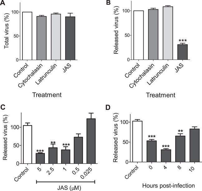
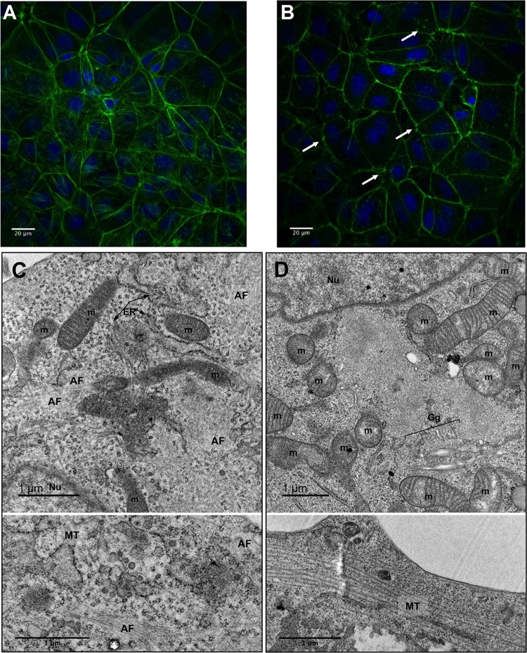
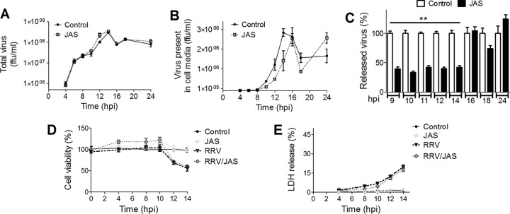
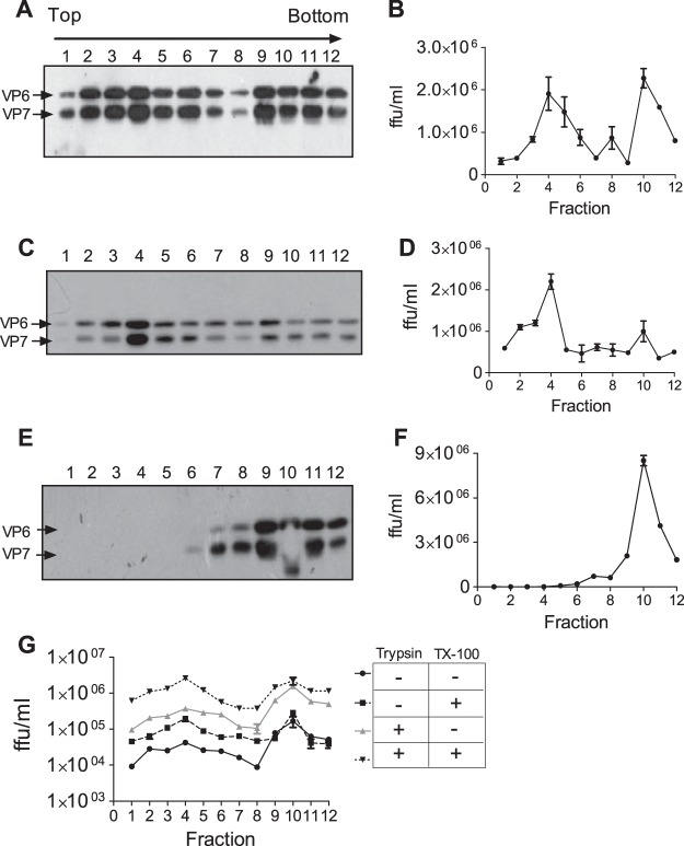
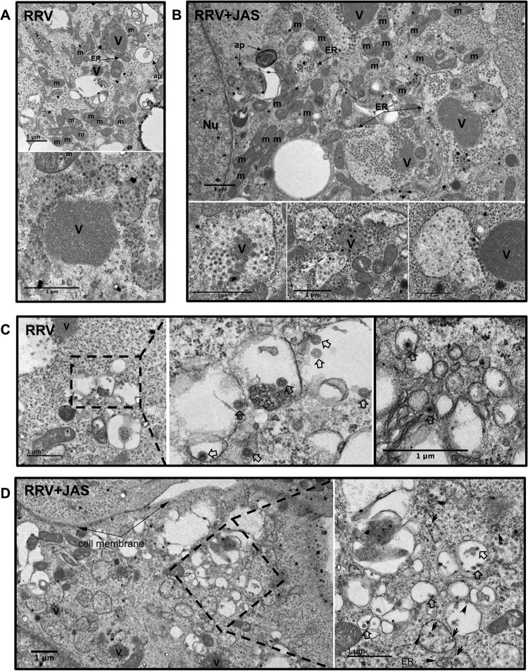
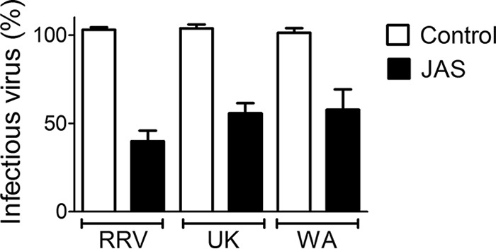
During the late stages of rotavirus morphogenesis, the surface proteins VP4 and VP7 are assembled onto the previously structured double-layered virus particles to yield a triple-layered, mature infectious virus. The current model for the assembly of the outer capsid is that it occurs within the lumen of the endoplasmic reticulum. However, it has been shown that VP4 and infectious virus associate with lipid rafts, suggesting that the final assembly of the rotavirus spike protein VP4 involves a post-endoplasmic reticulum event. In this work, we found that the actin inhibitor jasplakinolide blocks the cell egress of rotavirus from nonpolarized MA104 cells at early times of infection, when there is still no evidence of cell lysis. These findings contrast with the traditional assumption that rotavirus is released from nonpolarized cells by a nonspecific mechanism when the cell integrity is lost. Inspection of the virus present in the extracellular medium by use of density flotation gradients revealed that a fraction of the released virus is associated with low-density membranous structures. Furthermore, the intracellular localization of VP4, its interaction with lipid rafts, and its targeting to the cell surface were shown to be prevented by jasplakinolide, implying a role for actin in these processes. Finally, the VP4 present at the plasma membrane was shown to be incorporated into the extracellular infectious virus, suggesting the existence of a novel pathway for the assembly of the rotavirus spike protein.IMPORTANCE Rotavirus is a major etiological agent of infantile acute severe diarrhea. It is a nonenveloped virus formed by three concentric layers of protein. The early stages of rotavirus replication, including cell attachment and entry, synthesis and translation of viral mRNAs, replication of the genomic double-stranded RNA (dsRNA), and the assembly of double-layered viral particles, have been studied widely. However, the mechanisms involved in the later stages of infection, i.e., viral particle maturation and cell exit, are less well characterized. It has been assumed historically that rotavirus exits nonpolarized cells following cell lysis. In this work, we show that the virus exits cells by a nonlytic, actin-dependent mechanism, and most importantly, we describe that VP4, the spike protein of the virus, is present on the cell surface and is incorporated into mature, infectious virus, indicating a novel pathway for the assembly of this protein.

Keywords: cell exit; morphogenesis; rotavirus; virus assembly.


Figures

FIG 1
FIG 1
Release of rotavirus from MA104 cells decreases in the presence of JAS. MA104 cells were infected with RRV (MOI = 3). At 4 hpi, JAS (1 μM), latrunculin (1 μM), cytochalasin (10 mM), or DMSO (in the case of control cells) was added and kept in the medium; at 14 hpi, the amount of total infectious virus (virus present in the medium and cell-associated virus) (A) or virus present only in the medium (B) was quantitated as described in Materials and Methods. (C) Different concentrations of JAS or DMSO (in the case of control cells) were added after cell infection, and the drugs were kept in the medium; at 14 hpi, the virus present in the medium was quantitated. (D) JAS (1 μM) was added to infected cells at different times postinfection (0, 4, 8, and 10 h), and at 14 hpi, the amount of virus in the cell medium was quantitated as described above. Data are expressed as percentages of infectious virus present in the medium or of total virus compared to that in control cells treated with DMSO, which was taken as 100%. The arithmetic means and standard deviations for four independent experiments performed in triplicate are shown. **, P < 0.01; ***, P < 0.001.
FIG 2
FIG 2
Jasplakinolide affects the actin cytoskeleton structure. MA104 cells were left untreated (DMSO) (A) or treated with JAS (1 μM) (B) for 14 h at 37°C, fixed, immunostained, and analyzed by immunofluorescence assay. Actin filaments were stained with phalloidin coupled to Alexa 448 (green), and nuclei were stained with DAPI (blue). (C and D) Electron micrographs of MA104 cells that were left untreated (C) or treated with 0.5 μM JAS (D) for 4 h at 37°C. Cells were fixed and embedded as described in Materials and Methods. ER, endoplasmic reticulum; Gg, Golgi apparatus; m, mitochondria; Nu, nucleus; AF, actin filaments, MT, microtubules. The arrows indicate the ER membranes.
FIG 3
FIG 3
JAS affects the kinetics of rotavirus cell release. MA104 cells were infected with RRV (MOI = 3), either JAS (1 μM) or DMSO (control cells) was added at 4 hpi, and total virus (A) or the virus present in the extracellular medium (B) was quantitated at the indicated times as described in Materials and Methods. Data are expressed in FFU per milliliter. (C) Percentages of infectious virus present in the medium for JAS-treated cells compared to that for control cells treated with DMSO, which was taken as 100%. Infected or mock-infected cells, treated or not with JAS, were assayed for resazurin reduction (D) and LDH release (E) at the indicated times postinfection. The arithmetic means ± standard deviations for four independent experiments performed in triplicate are shown. **, P < 0.001.
FIG 4
FIG 4
A fraction of released infectious virus associates with low-density membranous structures. MA104 cells were infected with RRV (at an MOI of 3), and at 10 hpi the extracellular medium was collected. Viral particles present in the medium of untreated control cells (A), JAS-treated cells (C), or cells not treated with the drug but treated with 1% Triton X-100 (TX-100) for 30 min at room temperature (E) were fractionated by use of iodixanol gradients as described in Materials and Methods. Twelve 400-μl fractions were collected from the top of the tube, and the proteins present in each fraction were separated by SDS-PAGE (10% gel) and analyzed by Western blotting using anti-TLP antibodies. The viral titer of each fraction was quantitated as described in Materials and Methods, and titers are shown in panels B, D, and F. (G) Fractions from the gradient shown in panel A were left untreated or treated with 1% Triton X-100 for 30 min at room temperature and subsequently incubated or not with trypsin (10 μg/ml) for 30 min at 37°C, and the viral titer of each fraction was quantitated as described above. The arithmetic means ± standard deviations for three independent experiments are shown.
FIG 5
FIG 5
Cellular distribution of VP4 is affected by JAS. MA104 cells infected with RRV were treated or not with 1 μM JAS. The drug was added either at 0 hpi, in the case of cells that were fixed at 4 and 6 hpi, or at 4 hpi, for cells that were fixed at 10 hpi. VP4 was stained with MAb HS2 and goat anti-mouse IgG coupled to Alexa 448 (green). NSP2 was stained with a rabbit polyclonal antibody and a goat anti-rabbit antibody coupled to Alexa 568 (red). Nuclei, shown in blue, were stained with DAPI.
FIG 6
FIG 6
JAS disrupts the association between lipid rafts and viral particles. Infected MA104 cells were treated or not with JAS (1 μM). At 10 hpi, the cells were lysed and rafts were purified as described in Materials and Methods. (A) Eleven fractions were collected from each iodixanol gradient and analyzed by Western blotting using the anti-TLP antibody. As a raft marker, the presence of ganglioside GM1 was detected by dot blotting using the cholera toxin B subunit conjugated to horseradish peroxidase. (B) Infectivity assay. The titer of each fraction was determined on MA104 cells as described in Materials and Methods. Data are expressed as percentages of infectious virus present in the fractions from JAS-treated infected cells compared to that for infectious virus in each control fraction, which was taken as 100%. The arithmetic means and standard deviations for three independent experiments are shown. ***, P < 0.001.
FIG 7
FIG 7
Transport of VP4 to the plasma membrane is affected by JAS treatment. MA104 cells were infected with RRV (at an MOI of 3) and left untreated or treated with JAS (1 μM) for 6 h before being biotinylated. At the indicated times postinfection, the cells were biotinylated as described in Materials and Methods. The biotinylated proteins were precipitated by use of streptavidin (Strp) magnetic beads and analyzed by Western blotting using the anti-TLP antibody. The loading control was developed with a streptavidin-peroxidase conjugate. (A) Representative Western blot of proteins harvested at 6 hpi. A biotinylated membrane protein was used as a loading control. (B) Quantification of the biotinylated VP4 present at the plasma membrane by densitometry analysis as described in Materials and Methods. The amount of VP4 on the surfaces of control untreated cells at each time postinfection was taken as 100%. The arithmetic means and standard deviations for three independent experiments are shown. **, P < 0.01; ***, P < 0.001.
FIG 8
FIG 8
VP4 present at the plasma membrane is incorporated into viral particles. MA104 cells were infected with RRV (at an MOI of 3 FFU/cell), and at 6 hpi the cells were biotinylated with the impermeant reagent sulfo-NHS-LC-biotin for 30 min. The cells were then washed and incubated until 12 hpi. At this time, TLPs were purified twice by use of CsCl isopycnic gradients. (A) TLPs obtained from biotinylated (Biot) and nonbiotinylated (Ctrl) cells were purified by use of streptavidin (Strp) magnetic beads and analyzed by Western blotting using the anti-TLP antibody. (B) Control TLPs (1) and biotinylated TLPs (2) were stained with streptavidin-gold and analyzed by electron microscopy as described in Materials and Methods.
FIG 9
FIG 9
Electron microscopy analysis of MA104-infected cells in the presence of JAS. MA104 cells were infected with RRV at an MOI of 25 VFU per cell, and at 6 hpi the cells were left untreated (DMSO) (A) or treated with 0.5 μM JAS (B). At 10 hpi, the cells were fixed and embedded as described in Materials and Methods. (C and D) RRV-infected MA104 cells, as described above, in the absence (C) or presence (D) of 0.5 μM JAS. The dashed boxes show viral particles within vesicles close to the plasma membrane and far from viroplasms; in the right panels, higher-magnification views of the dashed boxes from the left panels are shown. ER, endoplasmic reticulum; m, mitochondria; Nu, nucleus; V, viroplasm. Also, viral particles in smooth vesicles (open arrows) and ER vesicles (black arrowheads) are shown.
FIG 10
FIG 10
Jasplakinolide affects the release of different rotavirus strains. MA104 cells were infected with the indicated rotavirus strains (at an MOI of 3 FFU/ml). At 12 hpi, the extracellular medium was collected and the released virus was quantified as described in Materials and Methods. Data are expressed as percentages of the amount of infectious virus present in the medium of control cells treated with DMSO, which was taken as 100%. The arithmetic means and standard deviations for three independent experiments performed in triplicate are shown.

Similar articles

Cited by

References

    1. Estes MK, Greenberg HB. 2013. Rotaviruses, p 1347–1401. In Knipe DM, Howley PM, Cohen JI, Griffin DE, Lamb RA, Martin MA, Racaniello VR, Roizman B (ed), Fields virology, 6th ed Lippincott Williams & Wilkins, Philadelphia, PA.
    1. Nejmeddine M, Trugnan G, Sapin C, Kohli E, Svensson L, Lopez S, Cohen J. 2000. Rotavirus spike protein VP4 is present at the plasma membrane and is associated with microtubules in infected cells. J Virol 74:3313–3320. doi:10.1128/JVI.74.7.3313-3320.2000. - DOI - PMC - PubMed
    1. Sapin C, Colard O, Delmas O, Tessier C, Breton M, Enouf V, Chwetzoff S, Ouanich J, Cohen J, Wolf C, Trugnan G. 2002. Rafts promote assembly and atypical targeting of a nonenveloped virus, rotavirus, in Caco-2 cells. J Virol 76:4591–4602. doi:10.1128/JVI.76.9.4591-4602.2002. - DOI - PMC - PubMed
    1. Enouf V, Chwetzoff S, Trugnan G, Cohen J. 2003. Interactions of rotavirus VP4 spike protein with the endosomal protein Rab5 and the prenylated Rab acceptor PRA1. J Virol 77:7041–7047. doi:10.1128/JVI.77.12.7041-7047.2003. - DOI - PMC - PubMed
    1. Li B, Ding S, Feng N, Mooney N, Ooi YS, Ren L, Diep J, Kelly MR, Yasukawa LL, Patton JT, Yamazaki H, Shirao T, Jackson PK, Greenberg HB. 2017. Drebrin restricts rotavirus entry by inhibiting dynamin-mediated endocytosis. Proc Natl Acad Sci U S A 114:E3642–E3651. doi:10.1073/pnas.1619266114. - DOI - PMC - PubMed

Publication types

LinkOut - more resources