. 2017 Dec 21;2(24):e96455.
doi: 10.1172/jci.insight.96455.

Eosinophils promote inducible NOS-mediated lung allograft acceptance

Affiliations

Eosinophils promote inducible NOS-mediated lung allograft acceptance

Oscar Okwudiri Onyema et al. JCI Insight. .

Abstract

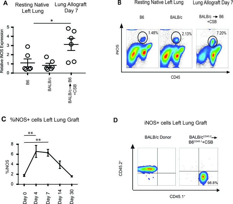
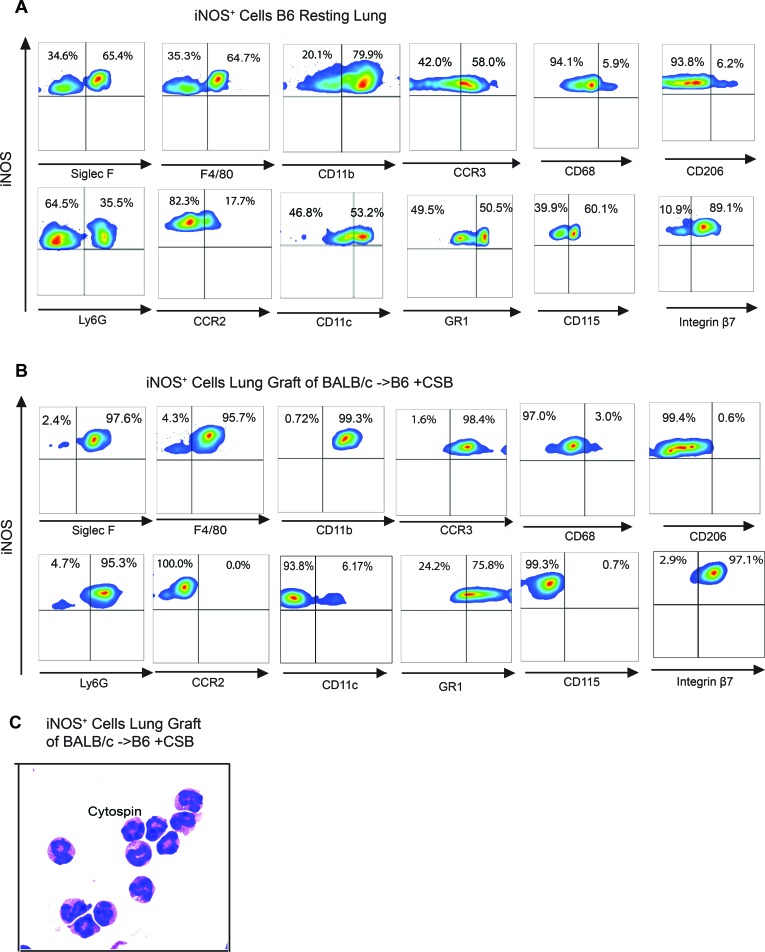
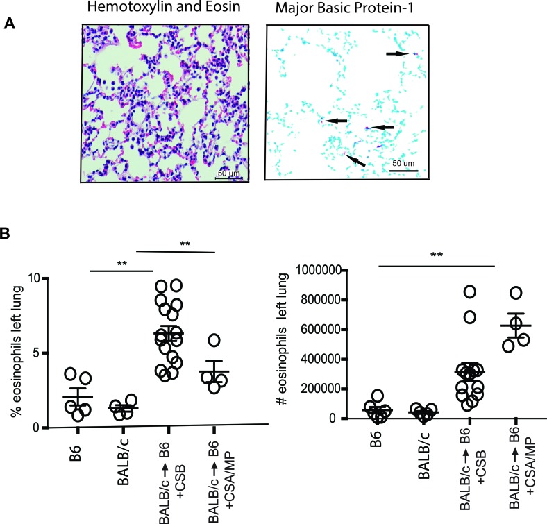
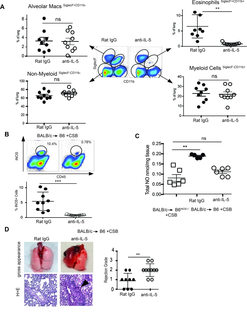
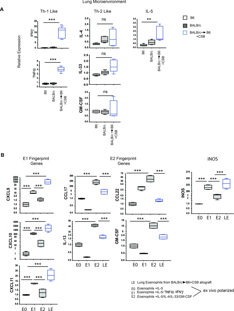
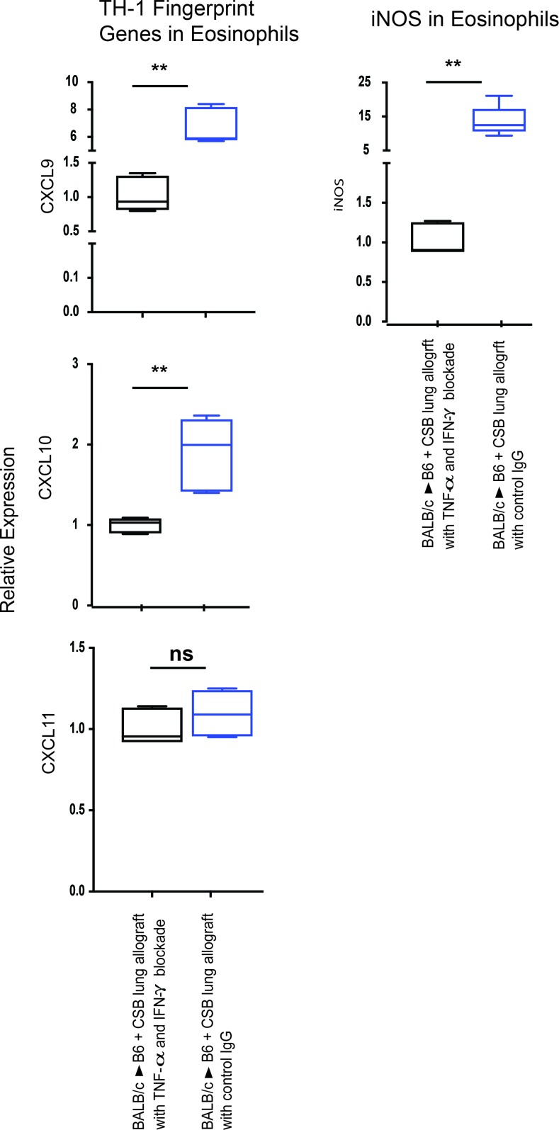
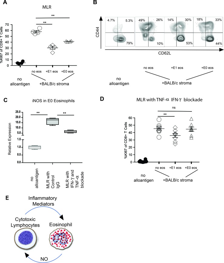
Lungs allografts have worse long-term survival compared with other organ transplants. This is most likely due to their unique immunoregulation that may not respond to traditional immunosuppression. For example, local NO generation by inducible NOS (iNOS) is critical for lung allograft acceptance but associates with rejection of other solid organs. The source of NO in accepting lung allografts remains unknown. Here, we report that, unlike the case for other pulmonary processes in which myeloid cells control NO generation, recipient-derived eosinophils play a critical and nonredundant role in iNOS-mediated lung allograft acceptance. Depletion of eosinophils reduces NO levels to that of recipients with global deletion of iNOS and leads to a costimulatory blockade-resistant form of rejection. Furthermore, NO production by eosinophils depends on Th1 polarization by inflammatory mediators, such as IFN-γ and TNF-α. Neutralization of such mediators abrogates eosinophil suppressive capacity. Our data point to what we believe to be a unique and previously unrecognized role of eosinophil polarization in mediating allograft tolerance and put into perspective the use of high-dose eosinophil-ablating corticosteroids after lung transplantation.

Keywords: Adaptive immunity; Immunology; Respiration; Th1 response; Transplantation.


Conflict of interest statement

Conflict of interest: The authors have declared that no conflict of interest exists.

Figures

Figure 1
Figure 1. Accepting lung allografts are infiltrated by recipient-derived iNOS+ cells.
(A) Quantitative RT-PCR revealed a significant increase in iNOS transcript levels 7 days after transplantation of BALB/c lungs to B6 recipients with CSB immunosuppression. (B) Flow cytometric analysis demonstrated that lung allografts are infiltrated by iNOS+ hematopoietic cells (representative of 8 separate transplants). (C) Time-course analysis demonstrated a gradual increase in iNOS+ cells, which decrease to baseline levels 1 month after transplantation (representative of at least 4 transplants per time point). (D) Transplantation of a BALB/cCD45.2+ lung grafts to a B6CD45.1+ recipient demonstrated that all iNOS+ graft-infiltrating cells are of recipient origin (representative of 3 separate transplants). *P <.05, **P < 0.01. Comparison by Friedman’s test was used for paired data, while the Kruskal-Wallis test was used for the unpaired group of observations. Post-hoc analysis of differences and comparison of differences between pairs of data were performed with the Wilcoxon rank test and the Mann-Whitney U test for paired and unpaired observations, respectively.
Figure 2
Figure 2. Phenotype of iNOS+ cells from resting lung and lung allografts.
(A) iNOS+ cells in resting B6 left lung are a heterogeneous population (representative of 7 transplants). (B) iNOS+ cells in BALB/c→B6 lung transplantation with CSB immunosuppression (representative of 7 transplants). (C) Cytospin preparation and Romanowsky staining of iNOS+ cells in accepting lung grafts, demonstrating an eosinophil phenotype. Original magnification, ×400.
Figure 3
Figure 3. Tissue distribution and quantification of eosinophils.
(A) Seven days after transplantation of BALB/c to B6 lung allografts with CSB immunosuppression eosinophils were distributed throughout the periphery of the lung in the alveolar space and septae, as indicated by the arrows. H&E staining and major basic protein-1 immunohistochemistry are shown. Scale bars: μm. (B) Quantitative analysis of eosinophils in resting B6 and BALB/c mice and BALB/c→B6 lung transplantation with CSB or CsA/MP immunosuppression as a percentage and total number of lung-resident cells. **P < 0.01. Comparison by Friedman’s test was used for paired data, while the Kruskal-Wallis test was used for the unpaired group of observations. Post-hoc analysis of differences and comparison of differences between pairs of data were performed with the Wilcoxon rank test and the Mann-Whitney U test for paired and unpaired observations, respectively.
Figure 4
Figure 4. Eosinophil depletion eliminates NO production and potentiates a CSB-resistant form of lung allograft rejection.
(A) Recipient IL-5 neutralization virtually eliminates all eosinophils in the lung allograft with minimal effect on other cell types (8–9 transplants per group). (B) IL-5 neutralization also eliminates iNOS-expressing graft-resident cells and (C) reduces lung graft NO production to levels statistically comparable to B6iNOS–/– recipients. (D) Depletion of eosinophils results in lung allograft rejection, as measured by gross changes, histologic evidence of inflammation, and ISHLT grade. Original magnification, ×100. ns, P > 0.05; **P < 0.01, ***P < 0.001. Comparison by Friedman’s test was used for paired data, while the Kruskal-Wallis test was used for the unpaired group of observations. Post-hoc analysis of differences and comparison of differences between pairs of data were performed with the Wilcoxon rank test and the Mann-Whitney U test for paired and unpaired observations, respectively.
Figure 5
Figure 5. Lung allograft acceptance is associated with Th1-like polarization.
(A) Gene expression analysis of lungs demonstrated a Th1-like polarization 4 days after transplantation (representative of 6 separate transplants, with BALB/c→B6 allografts outlined in blue, BALB/c resting lungs outlined in black with black dots, and B6 resting lungs outlined in black). (B) Eosinophils flow cytometrically sorted from BALB/c→B6 lung grafts 7 days after transplantation with CSB immunosuppression demonstrated an E1-like gene fingerprint pattern that resembles that of eosinophils exposed to IFN-γ and TNF-α ex vivo (data are from 5 separate transplants). **P < 0.01, ***P < 0.001. Comparison by Friedman’s test was used for paired data, while the Kruskal-Wallis test was used for the unpaired group of observations. Post-hoc analysis of differences and comparison of differences between pairs of data were performed with the Wilcoxon rank test and the Mann-Whitney U test for paired and unpaired observations, respectively. For the box plots, the bottom and top of the boxes represent the lower and upper quartiles, respectively; the dark band inside the box represents the median; and the top and bottom whiskers represent the maximum and minimum observed values, respectively.
Figure 6
Figure 6. Modulation of Th1 signature genes in lung graft eosinophils.
Expression of Th1 signature genes in lung graft eosinophils after IFN-γ and TNF-α blockade. Blue boxes represent eosinophils isolated from BALB/c→B6 transplanted lung grafts with CSB, and black boxes represent eosinophils isolated from BALB/c→B6 transplanted lung grafts with CSB in the presence of IFN-γ and TNF-α neutralization (representative of a minimum of 4 transplants per group). ns, P > 0.05; **P < 0.01. Comparison by Friedman’s test was used for paired data, while the Kruskal-Wallis test was used for the unpaired group of observations. Post-hoc analysis of differences and comparison of differences between pairs of data were performed with the Wilcoxon rank test and the Mann-Whitney U test for paired and unpaired observations, respectively. For the box plots, the bottom and top of the boxes represent the lower and upper quartiles, respectively; the dark band inside the box represents the median; and the top and bottom whiskers represent the maximum and minimum observed values, respectively.
Figure 7
Figure 7. Modulation of CD8+ T cell proliferation and differentiation by E1-polarized eosinophil.
(A) Proliferation, as measured by Ki67 expression among B6 CD45.1+ CD8+ T cells activated with BALB/c allogenic splenocytes, with or without the addition of E1 or E0 eosinophils. Data represent 1 of 2 experiments with 4 separate replicates per experiment. (B) In vitro differentiation, as measured by CD44 and CD62L expression on B6CD45.1+ CD8+ T cells stimulated by BALB/c allogenic splenocytes, with or without the addition of E1 or E0 eosinophils. (C) iNOS expression in CD8+ T cells activated with BALB/c allogenic splenocytes, with or without the addition of IFN-γ– and TNF-α–blocking antibodies (representative experiment of 4 separate MLRs). (D) Modulation of CD8+ T cell proliferation by E0 and E1 eosinophils in MLR of BALB/c allogenic splenocyte stimulators with IFN-γ and TNF-α blockade. (E) Schematic representation of the regulatory feedback loop between alloreactive lymphocytes and eosinophils in lung grafts. ns, P > 0.05; **P < 0.01. Comparison by Friedman’s test was used for paired data, while the Kruskal-Wallis test was used for the unpaired group of observations. Post-hoc analysis of differences and comparison of differences between pairs of data were performed with the Wilcoxon rank test and the Mann-Whitney U test for paired and unpaired observations, respectively. For the box plot, the bottom and top of the boxes represent the lower and upper quartiles, respectively; the dark band inside the box represents the median; and the top and bottom whiskers represent the maximum and minimum observed values, respectively.

References

    1. Conde P, et al. DC-SIGN(+) macrophages control the induction of transplantation tolerance. Immunity. 2015;42(6):1143–1158. doi: 10.1016/j.immuni.2015.05.009. - DOI - PMC - PubMed
    1. Garcia MR, et al. Monocytic suppressive cells mediate cardiovascular transplantation tolerance in mice. J Clin Invest. 2010;120(7):2486–2496. doi: 10.1172/JCI41628. - DOI - PMC - PubMed
    1. Li H, Shi B. Tolerogenic dendritic cells and their applications in transplantation. Cell Mol Immunol. 2015;12(1):24–30. doi: 10.1038/cmi.2014.52. - DOI - PMC - PubMed
    1. Kreisel D, et al. Emergency granulopoiesis promotes neutrophil-dendritic cell encounters that prevent mouse lung allograft acceptance. Blood. 2011;118(23):6172–6182. doi: 10.1182/blood-2011-04-347823. - DOI - PMC - PubMed
    1. Martinez OM, et al. Evidence for a nonclassical pathway of graft rejection involving interleukin 5 and eosinophils. Transplantation. 1993;55(4):909–918. doi: 10.1097/00007890-199304000-00041. - DOI - PubMed

Publication types