. 2017 Dec 20;7(1):17903.
doi: 10.1038/s41598-017-18224-x.

Interleukin-33 modulates inflammation in endometriosis

Affiliations

Interleukin-33 modulates inflammation in endometriosis

Jessica E Miller et al. Sci Rep. .

Abstract

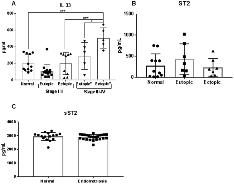
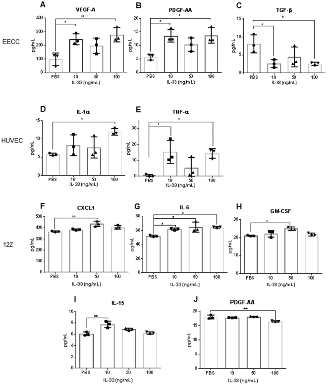
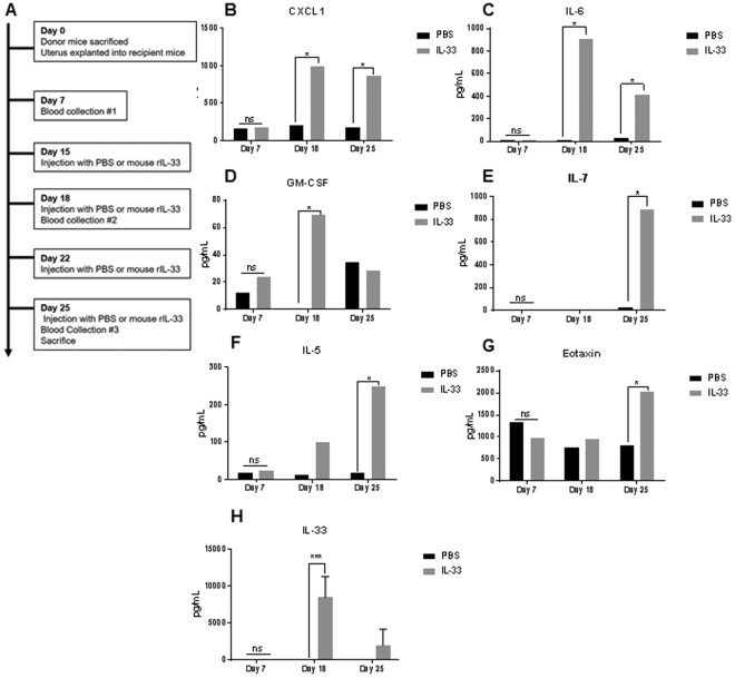
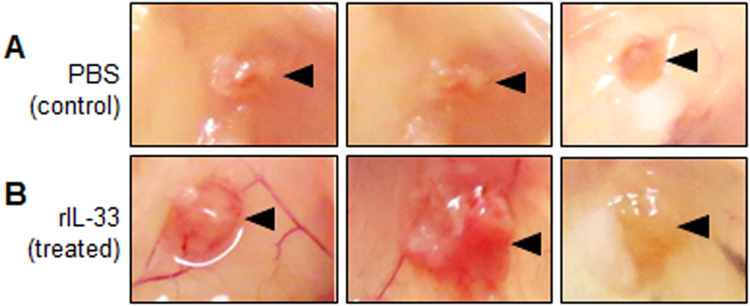
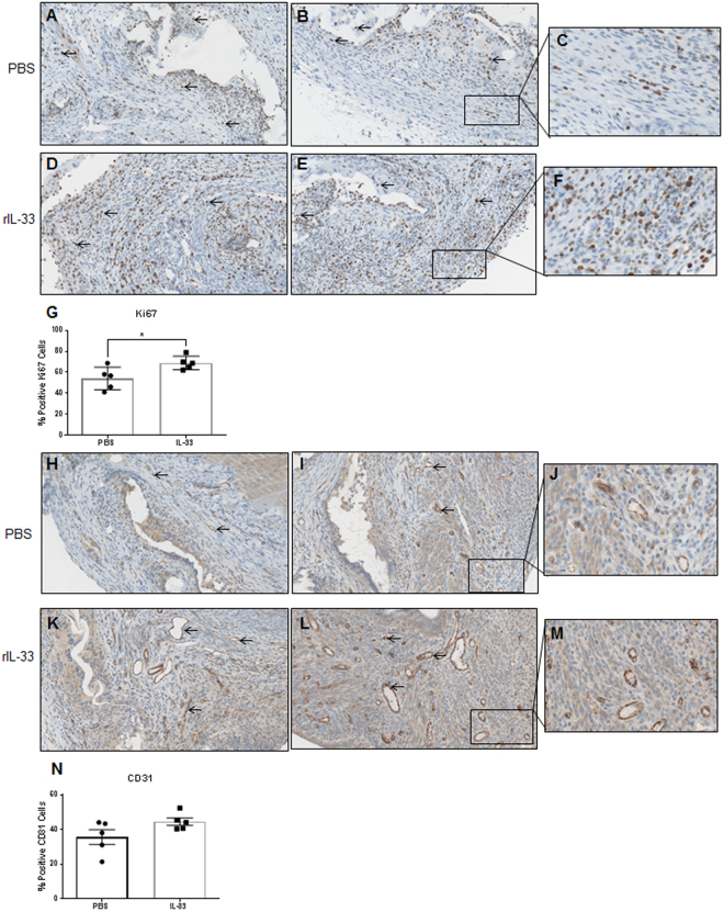
Endometriosis is a debilitating condition that is categorized by the abnormal growth of endometrial tissue outside the uterus. Although the pathogenesis of this disease remains unknown, it is well established that endometriosis patients exhibit immune dysfunction. Interleukin (IL)-33 is a danger signal that is a critical regulator of chronic inflammation. Although plasma and peritoneal fluid levels of IL-33 have been associated with deep infiltrating endometriosis, its contribution to the disease pathophysiology is unknown. We investigated the role of IL-33 in the pathology of endometriosis using patient samples, cell lines and a syngeneic mouse model. We found that endometriotic lesions produce significantly higher levels of IL-33 compared to the endometrium of healthy, fertile controls. In vitro stimulation of endometrial epithelial, endothelial and endometriotic epithelial cells with IL-33 led to the production of pro-inflammatory and angiogenic cytokines. In a syngeneic mouse model of endometriosis, IL-33 injections caused systemic inflammation, which manifested as an increase in plasma pro-inflammatory cytokines compared to control mice. Furthermore, endometriotic lesions from IL-33 treated mice were highly vascularized and exhibited increased proliferation. Collectively, we provide convincing evidence that IL-33 perpetuates inflammation, angiogenesis and lesion proliferation, which are critical events in the lesion survival and progression of endometriosis.


Conflict of interest statement

The authors declare that they have no competing interests.

Figures

Figure 1
Figure 1
Protein levels of IL-33, ST2 and sST2 in plasma and tissue samples from healthy, fertile volunteers and endometriosis patients. (A) Levels of IL-33 are significantly higher in the ectopic tissue from advanced staged endometriosis patients (n = 5) compared to healthy fertile controls (n = 11). No significant differences were found between healthy, fertile controls compared to early staged patients (n = 14) or eutopic tissue from advanced staged patients (n = 5). (B) The ST2 receptor is expressed on ectopic, endometriotic tissue. No significant differences were found between levels of ST2 in matched tissue from endometriosis patients (n = 7) compared to healthy, fertile controls (n = 11). (C) No significant differences were found between levels of sST2 in the plasma of endometriosis patients (n = 24) compared to healthy, fertile controls (n = 16) *p < 0.05 **p < 0.01 ***p < 0.0001.
Figure 2
Figure 2
Cell supernatant cytokine profile in EECC, HUVEC and 12Z upon stimulation with varying concentrations of human rIL-33 (10, 50 and 100 ng/mL). All experiments were conducted in triplicates. Cell supernatant was analyzed using a human multiplex assay and non-significant cytokines are not shown. (AC) Endometrial epithelial carcinoma cells (EECC) produced significantly higher levels of VEGF and PDGF-AA and a significant reduction in the level of TGF-b. (C,D) Human umbilical vein endothelial cells (HUVEC) produced significantly higher levels of IL-1a and TNF-a. (FJ) Endometriotic epithelial cells (12Z) produced significantly higher levels of CXCL1, IL-15, GM-CSF and IL-6 and significantly lower levels of PDGF-AA. *p < 0.05 **p < 0.01 ***p < 0.0001.
Figure 3
Figure 3
Plasma cytokine profile in mice with endometriosis and treated with PBS (control) (n = 5) or mouse rIL-33 (treated) (n = 5). Plasma was analyzed using a mouse multiplex assay and non-significant cytokines are not shown. (A) An outline of mouse experiment. (BG) Plasma cytokines revealed significantly higher levels of Eotaxin, GM-CSF, IL-6, IL-7, IL-5, CXCL1, and IL-33. *p < 0.05 **p < 0.01 ***p < 0.0001.
Figure 4
Figure 4
Endometriotic lesions harvested from PBS (control) and mouse rIL-33 (treated) mice. Endometriotic lesions harvested from mouse r-IL33 mice (n = 5) appear qualitatively larger with enhanced vasculature (B) compared to PBS treated (n = 5) mice (A). Black triangles indicate the endometriotic lesion.
Figure 5
Figure 5
Immunohistochemistry with a proliferation marker, Ki67 (AF) and vasculature marker CD31 (HM) revealed intense immunostaining. Semi-quantitative analysis of Ki67 (G) showed significant increase in % positive Ki67 cells in the mouse rIL-33 treated mice compared to control. Semi-quantitative analysis of CD31 (N) immunostaining shows a higher trend in rIL33 treated mice compared to controls. Black arrows indicate positive staining. *p < 0.05 **p < 0.01 ***p < 0.0001.

References

    1. Halme J, Hammond MG, Hulka JF, Raj SG, Talbert LM. Retrograde menstruation in healthy women and in patients with endometriosis. Obstet. 1984;64:151–154. - PubMed
    1. Ahn, S. H. et al. Pathophysiology and immune dysfunction in endometriosis. BioMed Research International2015 (2015). - PMC - PubMed
    1. Miller JE, et al. Implications of immune dysfunction on endometriosis associated infertility. Oncotarget. 2016 - PMC - PubMed
    1. Kyama CM, Debrock S, Mwenda JM, D’Hooghe TM. Potential involvement of the immune system in the development of endometriosis. Reprod. Biol. Endocrinol. 2003;1:123. doi: 10.1186/1477-7827-1-123. - DOI - PMC - PubMed
    1. Monsanto SP, et al. Surgical removal of endometriotic lesions alters local and systemic proinflammatory cytokines in endometriosis patients. Fertil. Steril. 2016;105:968–977.e5. doi: 10.1016/j.fertnstert.2015.11.047. - DOI - PMC - PubMed

Publication types

MeSH terms