. 2017 Dec 20;7(1):17892.
doi: 10.1038/s41598-017-17917-7.

Regulation of ribonucleotide synthesis by the Pseudomonas aeruginosa two-component system AlgR in response to oxidative stress

Affiliations

Regulation of ribonucleotide synthesis by the Pseudomonas aeruginosa two-component system AlgR in response to oxidative stress

Anna Crespo et al. Sci Rep. .

Abstract

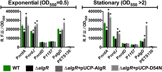
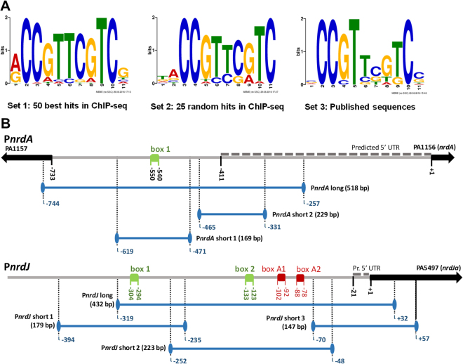
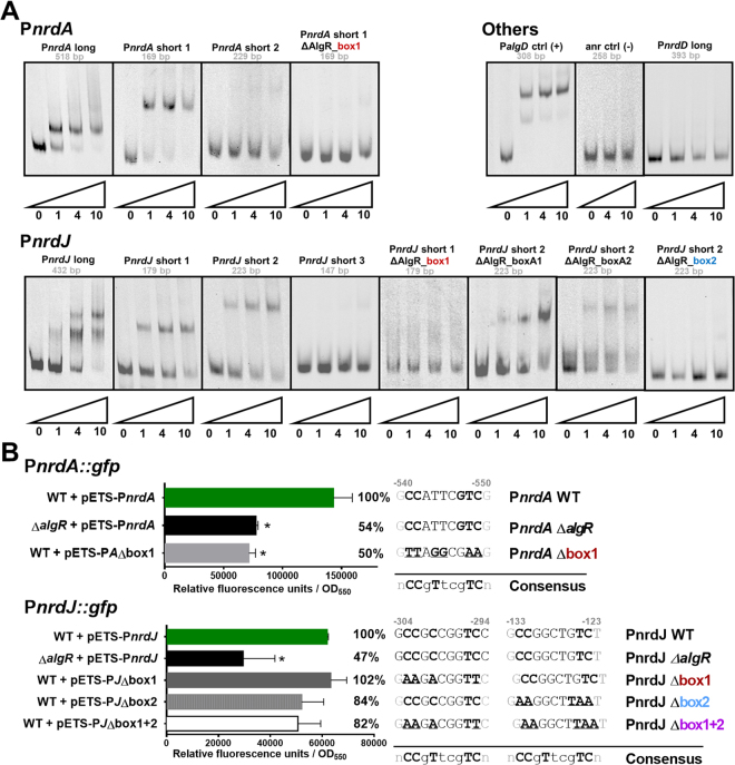
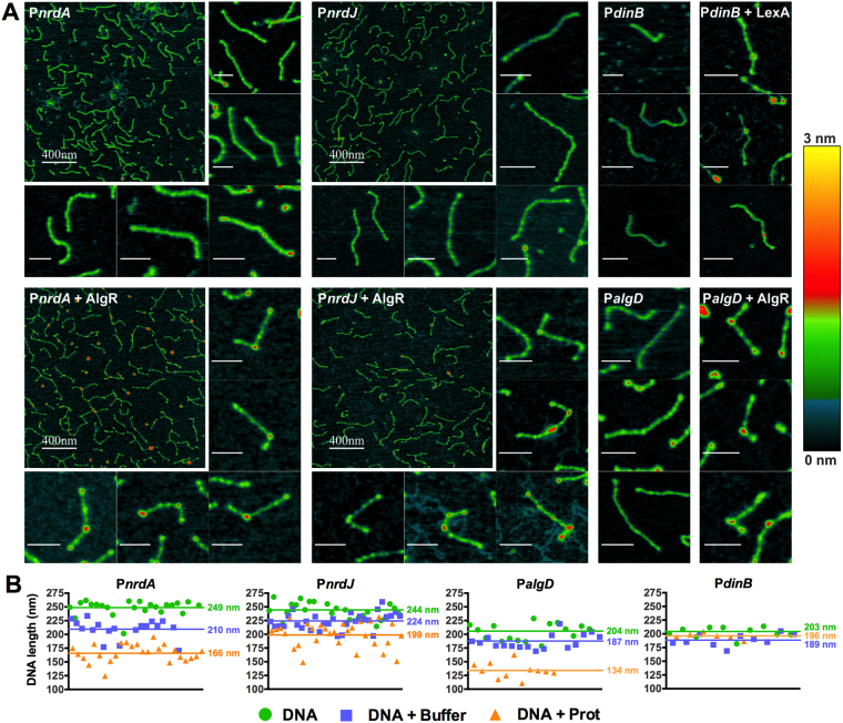
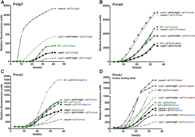
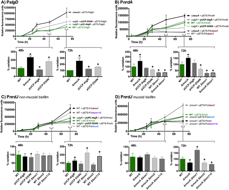
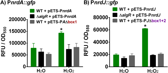
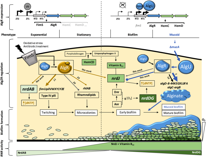
Ribonucleotide reductases (RNR) catalyze the last step of deoxyribonucleotide synthesis, and are therefore essential to DNA-based life. Three forms of RNR exist: classes I, II, and III. While eukaryotic cells use only class Ia RNR, bacteria can harbor any combination of classes, granting them adaptability. The opportunistic pathogen Pseudomonas aeruginosa surprisingly encodes all three classes, allowing it to thrive in different environments. Here we study an aspect of the complex RNR regulation whose molecular mechanism has never been elucidated, the well-described induction through oxidative stress, and link it to the AlgZR two-component system, the primary regulator of the mucoid phenotype. Through bioinformatics, we identify AlgR binding locations in RNR promoters, which we characterize functionally through EMSA and physically through AFM imaging. Gene reporter assays in different growth models are used to study the AlgZR-mediated control on the RNR network under various environmental conditions and physiological states. Thereby, we show that the two-component system AlgZR, which is crucial for bacterial conversion to the mucoid phenotype associated with chronic disease, controls the RNR network and directs how the DNA synthesis pathway is modulated in mucoid and non-mucoid biofilms, allowing it to respond to oxidative stress.


Conflict of interest statement

The authors declare that they have no competing interests.

Figures

Figure 1
Figure 1
In vivo AlgR regulation of RNR promoters and related genes. Gene reporter assays for PnrdA (pETS134), PnrdJ (pETS180), PnrdD (pETS136), PPA1157 (pETS206) and PalgD (pETS205) fused to GFP at exponential and stationary growth phases. Values are averages from at least three independent experiments, and error bars show positive standard deviation. The promoterless pETS130-GFP plasmid values are provided for comparison. Asterisks (*) indicate a statistically significant difference from the wild-type strain (p-value less than 0.05 in pairwise t-tests). Shortened names are used (see Supplementary Table S1).
Figure 2
Figure 2
AlgR boxes in the RNR promoters. (A) Sequence logos for the AlgR binding box. HMM logos are generated from count matrices (see Supplementary Fig. S1) produced by FIMO using three different sets of sequences containing AlgR binding sites (see Materials and Methods). (B) Schematics for promoters PnrdA (RNR class I promoter) and PnrdJ (RNR class II promoter). Identified boxes are represented in green, artifact boxes identified as false positives in the bioinformatics search are represented in red. Genes are represented by arrows; gene znuA has been eliminated from the PnrdJ schematic for improved readability. An approximated prediction of the 5′UTR for the studied operons (BPROM) is shown as dashed lines. Locations are indicated in base pairs relative to the ATG translation start codon of the first gene of the corresponding operon. DNA probes used for EMSA studies are indicated by solid blue lines.
Figure 3
Figure 3
Functional study of the AlgR boxes in RNR promoters. (A) EMSA experiments promoters PnrdA, PnrdJ, and PnrdD, together with positive control (PalgD) and negative control probes. Probe sizes are indicated below their names; numbers below the triangles represent pmol of AlgR. (B) Gene reporter assay for PnrdA and PnrdJ, during the early stationary phase (OD550 = 2.0) and under aerobic conditions. Error bars represent positive standard deviations; the asterisk indicates a statistically significant difference from the wild-type strain (p-value less than 0.05 in pairwise t-tests). The exact mutations introduced are detailed at the right of the graphic, and a simplified consensus sequence of the AlgR box is provided for comparison. The position of each box is indicated in bp (to the ATG of the first gene in the operon). Shortened names are used (see Supplementary Table S1). The images in A were cropped for clarity from the originals in Supplementary Fig. 2.
Figure 4
Figure 4
Atomic force microscopy images of DNA and DNA-protein complexes. (A) AFM images of DNA molecules or DNA-protein complexes, taken on mica under ambient conditions, are shown for PnrdA, PnrdJ, PalgD and PdinB promoters. Small images depict single DNA probes; scale bars without numbers above represent 80 nm. For the PnrdA and PnrdJ promoters, a general image at a higher scale is also shown. Colors represent the height of the structures, according to the scale at the right. (B) comparison of the apparent length of randomly selected units of all DNA probes when, before drying, they were in water (images not shown) or in binding buffer, as well as when complexed with AlgR.
Figure 5
Figure 5
AlgR regulation of RNR promoters during surface colonization. GFP-based gene reporter assays for PalgD (pETS205), (A) PnrdA (pETS134), (B) and PnrdJ (pETS180), (C) promoters fused to GFP, during surface colonization. GFP fluorescence is measured at different times of incubation during colony formation and presented as relative fluorescence units. Mucoid strains (PAOMA, PAO ΔmucA) are included. A fourth panel (D) shows further experiments with PnrdJ AlgR boxes to study the fine regulation performed at this level. For improved readability, shortened names are used (see Supplementary Table S1), the key features of each strain are highlighted in bold, wild-type strains are underlined and mutant boxes are color-coded.
Figure 6
Figure 6
AlgR regulation of RNR promoters in mucoid and non-mucoid biofilms. Gene reporter assay at different time points during static biofilm formation for PalgD (pETS205), (A), PnrdA (pETS134), (B) and PnrdJ (pETS180), (C and D). The values shown are the means of three independent experiments in 8 wells; error bars indicate positive and negative standard deviation. Shortened names are used (see Supplementary Table S1). For 48 h and 72 h, results are depicted as bar graphs; error bars show positive standard deviation, and the asterisk indicates a statistically significant difference from the wild-type strain (p-value less than 0.05 in pairwise t-tests).
Figure 7
Figure 7
AlgR regulation of RNR during oxidative stress. Gene reporter assays for the PnrdA and PnrdJ promoters fused to GFP. All strains were grown to OD550 = 0.5 and then subjected to 30 minutes of incubation with a stressing agent (1.0 mM H2O2) or control (equivalent volume of water). Values are averages from three independent experiments, and error bars show positive standard deviation. Asterisks (*) indicate statistically significant difference from the untreated wild-type strain (p-value less than 0.05 in pairwise t-tests). Shortened names are used (see Supplementary Table S1).
Figure 8
Figure 8
Model of AlgZR regulation of ribonucleotide reduction. Schematic representation of the AlgZR regulation of ribonucleotide reduction, in the context of other AlgZR-mediated regulation events. Arrows represent positive regulation, while lines with a bullet point represent negative regulation. Events characteristic of phosphorylated AlgR are highlighted in yellow, and events happening mostly in the mucoid phenotype are highlighted in blue. The representation is not exhaustive, and some events are eliminated for the sake of clarity. The source of information for any AlgR regulative event is indicated beside the corresponding line.

Similar articles

Cited by

References

    1. Govan JR, Deretic V. Microbial pathogenesis in cystic fibrosis: mucoid Pseudomonas aeruginosa and Burkholderia cepacia. Microbiol Rev. 1996;60:539–574. - PMC - PubMed
    1. Deretic V, Dikshit R, Konyecsni WM, Chakrabarty AM, Misra TK. The algR gene, which regulates mucoidy in Pseudomonas aeruginosa, belongs to a class of environmentally responsive genes. J Bacteriol. 1989;171:1278–1283. doi: 10.1128/jb.171.3.1278-1283.1989. - DOI - PMC - PubMed
    1. Mohr CD, Hibler NS, Deretic V. AlgR, a response regulator controlling mucoidy in Pseudomonas aeruginosa, binds to the FUS sites of the algD promoter located unusually far upstream from the mRNA start site. J Bacteriol. 1991;173:5136–5143. doi: 10.1128/jb.173.16.5136-5143.1991. - DOI - PMC - PubMed
    1. Leid JG, et al. The exopolysaccharide alginate protects Pseudomonas aeruginosa biofilm bacteria from IFN-gamma-mediated macrophage killing. J Immunol. 2005;175:7512–7518. doi: 10.4049/jimmunol.175.11.7512. - DOI - PubMed
    1. Hentzer M, et al. Alginate overproduction affects Pseudomonas aeruginosa biofilm structure and function. J Bacteriol. 2001;183:5395–5401. doi: 10.1128/JB.183.18.5395-5401.2001. - DOI - PMC - PubMed

Publication types

MeSH terms