Case Reports
. 2017 Jul 10:2:23.
doi: 10.1038/s41525-017-0024-5. eCollection 2017.

A homozygous mutation in the stem II domain of RNU4ATAC causes typical Roifman syndrome

Affiliations
Case Reports

A homozygous mutation in the stem II domain of RNU4ATAC causes typical Roifman syndrome

Yael Dinur Schejter et al. NPJ Genom Med. .

Abstract

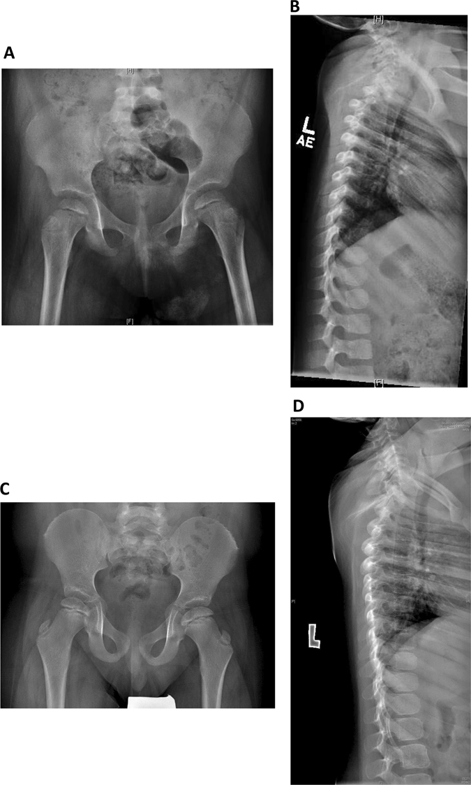
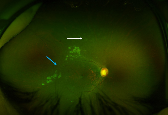
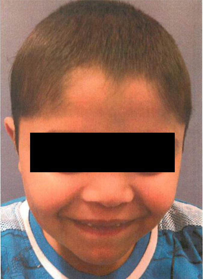
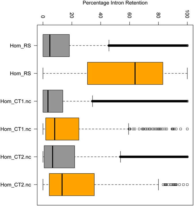
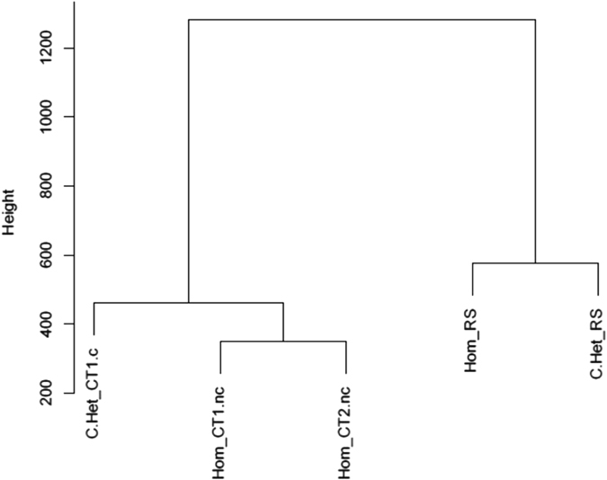
Roifman syndrome (OMIM# 616651) is a complex syndrome encompassing skeletal dysplasia, immunodeficiency, retinal dystrophy and developmental delay, and is caused by compound heterozygous mutations involving the Stem II region and one of the other domains of the RNU4ATAC gene. This small nuclear RNA gene is essential for minor intron splicing. The Canadian Centre for Primary Immunodeficiency Registry and Repository were used to derive patient information as well as tissues. Utilising RNA sequencing methodologies, we analysed samples from patients with Roifman syndrome and assessed intron retention. We demonstrate that a homozygous mutation in Stem II is sufficient to cause the full spectrum of features associated with typical Roifman syndrome. Further, we demonstrate the same pattern of aberration in minor intron retention as found in cases with compound heterozygous mutations.


Conflict of interest statement

The authors declare that they have no competing financial interests.Medical records were compiled into the Canadian Centre for Primary Immunodeficiency Registry, which has been approved by the Sick Kids Research Ethics board (protocol number 1000005598). This includes consent and assent from patients and family members for genetics analysis.

Figures

Fig. 1
Fig. 1
RNU4ATAC structural elements, and MOPD1 and Roifman syndrome causal variants. Stem I and Stem II are both elements at the 3′ and 5′ of U4atac, respectively, which base pair with U6atac. These elements are separated by an intramolecular 5′ stem-loop. Another stem loop is located at the 3′ end of U4atac. The Sm protein binding site is important for binding of the Sm proteins, which are important for the assembly of the complex and its import into the nucleus. The Stem II, Stem I, 5′ stem-loop and Sm binding site are all highly conserved. Delineated in red is the Stem II domain, which is obligatory for the pathogenesis of Roifman syndrome. Adapted from refs. ,
Fig. 2
Fig. 2
Skeletal features of Roifman syndrome in patients 1 and 2. a Pelvic X-ray of patient 1 featuring flattening of the humoral heads and shortening of the femoral necks, representing early stages of spondyloepiphyseal skeletal dysplasia. b Lateral spine X-ray of patient 1 featuring anterior vertebral notching of the lower thoracic vertebrae, and loss of lumbar lordosis. c Pelvic X-ray of patient 2 featuring bilateral small, flattened and slightly broadened femoral heads. d Lateral spine X-ray of patient 2 featuring loss of lumbar lordosis.
Fig. 3
Fig. 3
Retinal features of Roifman syndrome in patient 1. Retinal exam featuring normal optic discs, dull foveal reflex, attenuated retinal vessels (blue arrow) and background retinal pigment epithelial changes in the middle and far periphery (white arrow).
Fig. 4
Fig. 4
Facial features of the patient 2. Narrow palpebral fissures, a long philtrum and a thin upper lip are evident, in keeping with the classical facial features of Roifman syndrome patients. Informed consent was obtained for publication of patient images.
Fig. 5
Fig. 5
Pedigree of Roifman syndrome patients. a Pedigree of kindred 1 (K1), the family of patient 1, showing a c.16 G > A homozygous mutation. b Sequencing analysis of patient 1 and her heterozygous carrier parents. c Pedigree of kindred 2 (K2), the family of patient 2, showing a compound heterozygous mutation c.116 A > G/17 G > A. d Sequencing analysis of patient 2 and his heterozygous carrier parents. e Sequencing analysis of wild type RNU4ATAC. Circles represent female subjects, while squares denote male subjects. The black colour represents an affected status. The grey colour represents individuals who were unavailable for sequencing. The white colour represents affected non carrier, and half black–half white colour represents non affected heterozygous carrier. [=] indicates no variant detected.
Fig. 6
Fig. 6
Percentage intron retention (PIR) in patient 1 and unaffected family members. PIR across different introns is displayed as a boxplot: the affected patient 1 clearly displays greater minor intron retention than the unaffected wild-type siblings, whereas major intron retention levels are lower and similar across samples. Hom_RS-patient 1; Hom_CT1.nc-unaffected, non-carrier male sibling of patient 1, aged 3 months; Hom_CT2.nc-unaffected, non-carrier male sibling of patient 1, aged 9 years. Grey boxes–major intron retention levels, orange boxes–minor intron retention levels.
Fig. 7
Fig. 7
Cluster analysis for minor intron retention of affected subjects vs. controls. Cluster analysis was performed using the euclidean distance metrics. A clear discrimination of minor intron retention levels is demonstrated between affected subjects of both heterozygous and homozygous mutations, and their unaffected controls. C.Het_CT1.c-Unaffected carrier mother of patient 2; Hom_CT1.nc-unaffected, non-carrier male sibling of patient 1, aged 3 months; Hom_CT2.nc-unaffected, non-carrier male sibling of patient 1, aged 9 years; Hom_RS-Patient 1; C.Het_RS-Patient 2.

References

    1. Roifman CM. Immunological aspects of a novel immunodeficiency syndrome that includes antibody deficiency with normal immunoglobulins, spondyloepiphyseal dysplasia, growth and developmental delay, and retinal dystrophy. Can. J. Allergy Clini. Immunol. 1997;2:94–98.
    1. Roifman CM. Antibody deficiency, growth retardation, spondyloepiphyseal dysplasia and retinal dystrophy: a novel syndrome. Clin. Genet. 1999;55:103–9. doi: 10.1034/j.1399-0004.1999.550206.x. - DOI - PubMed
    1. Mandel K, Grunebaum E, Benson L. Noncompaction of the myocardium associated with Roifman syndrome. Cardiol. Young. 2001;11:240–3. doi: 10.1017/S1047951101000208. - DOI - PubMed
    1. Robertson SP, Rodda C, Bankier A. Hypogonadotrophic hypogonadism in Roifman syndrome. Clin. Genet. 2000;57:435–8. doi: 10.1034/j.1399-0004.2000.570606.x. - DOI - PubMed
    1. de Vries PJ, McCartney DL, McCartney E, Woolf D, Wozencroft D. The cognitive and behavioural phenotype of Roifman syndrome. J. Intellect. Disabil. Res. 2006;50:690–6. doi: 10.1111/j.1365-2788.2006.00817.x. - DOI - PubMed

Publication types

LinkOut - more resources