. 2017 Nov 1:2:34.
doi: 10.1038/s41525-017-0034-3. eCollection 2017.

Workflow optimization of whole genome amplification and targeted panel sequencing for CTC mutation detection

Affiliations

Workflow optimization of whole genome amplification and targeted panel sequencing for CTC mutation detection

Haiyan E Liu et al. NPJ Genom Med. .

Abstract

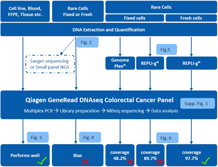
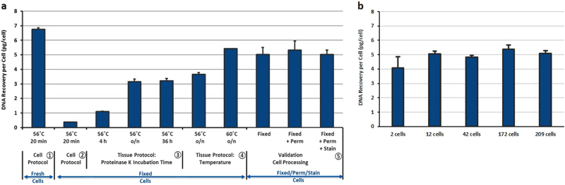
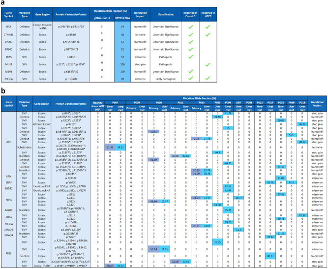
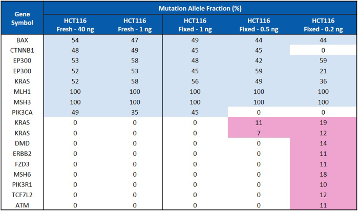
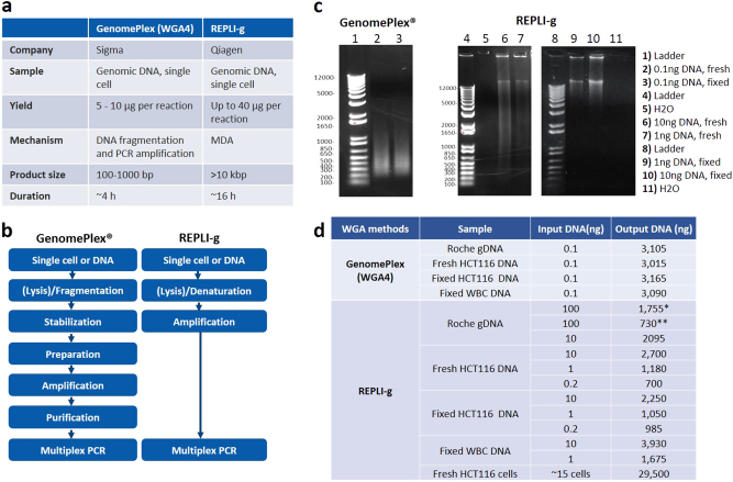
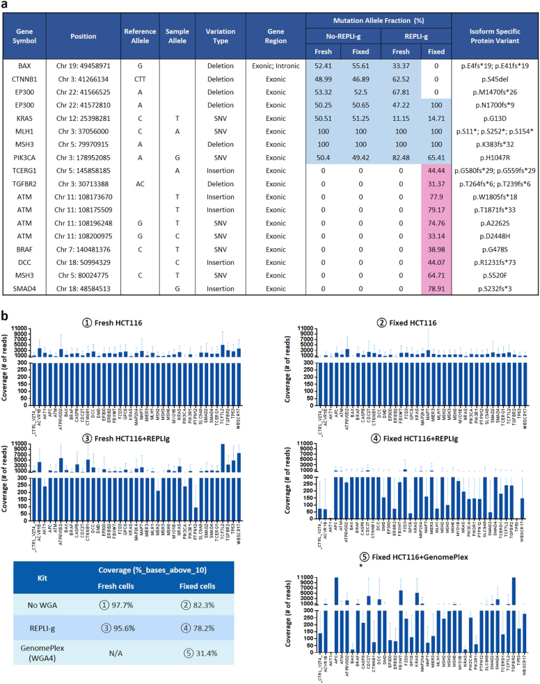
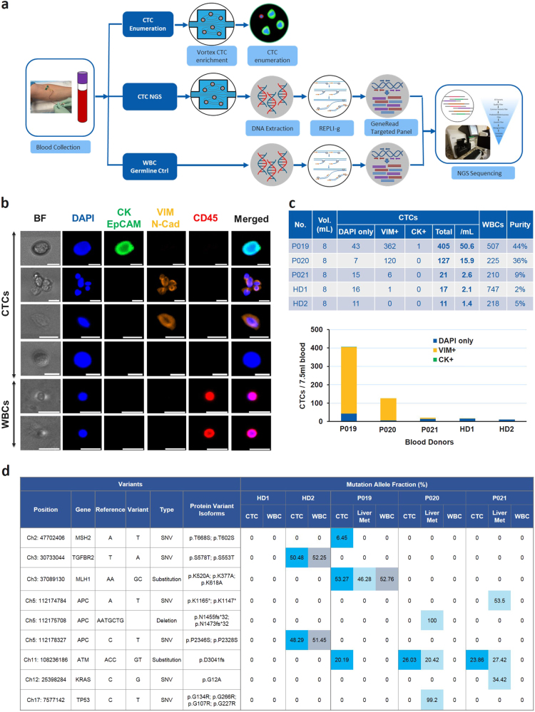
Genomic characterization of circulating tumor cells (CTCs) may prove useful as a surrogate for conventional tissue biopsies. This is particularly important as studies have shown different mutational profiles between CTCs and ctDNA in some tumor subtypes. However, isolating rare CTCs from whole blood has significant hurdles. Very limited DNA quantities often can't meet NGS requirements without whole genome amplification (WGA). Moreover, white blood cells (WBC) germline contamination may confound CTC somatic mutation analyses. Thus, a good CTC enrichment platform with an efficient WGA and NGS workflow are needed. Here, Vortex label-free CTC enrichment platform was used to capture CTCs. DNA extraction was optimized, WGA evaluated and targeted NGS tested. We used metastatic colorectal cancer (CRC) as the clinical target, HCT116 as the corresponding cell line, GenomePlex® and REPLI-g as the WGA methods, GeneRead DNAseq Human CRC Panel as the 38 gene panel. The workflow was further validated on metastatic CRC patient samples, assaying both tumor and CTCs. WBCs from the same patients were included to eliminate germline contaminations. The described workflow performed well on samples with sufficient DNA, but showed bias for rare cells with limited DNA input. REPLI-g provided an unbiased amplification on fresh rare cells, enabling an accurate variant calling using the targeted NGS. Somatic variants were detected in patient CTCs and not found in age matched healthy donors. This demonstrates the feasibility of a simple workflow for clinically relevant monitoring of tumor genetics in real time and over the course of a patient's therapy using CTCs.


Conflict of interest statement

Some of the authors (H.E.L., M.V., J.C., C.R., E.S.C.) have financial interests in Vortex Biosciences and intellectual property described herein. All the other authors declare that they have no competing financial interests.

Figures

Fig. 1
Fig. 1
Workflow optimization for biopsy genomic profiling, tissue and CTCs. Qiagen QIAamp Micro Kit and GeneRead DNAseq targeted panel sequencing were applied on all types of cell samples. This overall workflow performs well when DNA amount is sufficient, i.e., from whole blood and tissue biopsy. For rare cells, DNA input is insufficient for targeted NGS, leading to a bias and the need for an extra step of Whole Genome Amplification (WGA). Two WGA kits were evaluated (WGA4 from Sigma and REPLI-g from Qiagen) and obtained low coverage for fixed cells, while REPLI-g was identified as optimal for fresh rare cells and used for a final validation on patient CTCs
Fig. 2
Fig. 2
a Optimization of DNA extraction from rare and fixed cells using Qiagen QIAamp DNA Micro Kit. ~200–300 cells/experiment, N ≥ 2 per condition. ① Fresh cells using Cell Protocol yielded 6–7 pg of DNA/cell and were used as a control. ② Cell Protocol did not work on cells fixed with 4% PFA. ③ Tissue Protocol significantly increased the DNA yield when Proteinase K digestion was extended from 4 to 36 h, with significant advantage of overnight incubation. ④ Increasing the Proteinase K digestion temperature from 56 to 60 °C further increased the DNA yield from 60 to 80%. ⑤ The optimized protocol was then verified on fixed, fixed+permeabilized and fixed+permeabilized+stained HCT116 cells to mimic the output from CTC enrichment platforms. b Validation of an optimal DNA extraction protocol for low number of fixed cells. Different amounts of HCT116 cells fixed with 4% PFA were seeded inside a 96 well-plate, imaged and counted. DNA was extracted from these cells using the optimized protocol defined in A (Qiagen QIAamp DNA Micro Kit, Tissue Protocol, Proteinase K digestion overnight at 60 °C) and quantitated using qPCR
Fig. 3
Fig. 3
Mutation detection using GeneRead DNAseq targeted CRC panel sequencing. a Mutation detection in HCT116 cancer cells. Forty nanogram of DNA extracted from fresh HCT116 cells were subjected to multiplex PCR, library preparation and MiSeq sequencing. Mutations and corresponding allele frequencies were called using Ingenuity variant analysis software and compared to results reported from Cosmic* (catalog of somatic mutations in cancer) and ATCC websites. b Mutation detection in CRC patient biopsy tissues. Forty nanogram of DNA extracted from primary tumor and liver metastasis were subjected to multiplex PCR, library preparation and MiSeq sequencing. Mutations and corresponding Mutation Allele Fraction (MAF) were analyzed and compared between primary tumor and liver metastasis. In both tables, the blue color highlights the detected mutation and the number inside each cell represents the MAF of the mutation
Fig. 4
Fig. 4
Mutation detection in HCT116 cancer cells with different DNA input using GeneRead DNAseq targeted CRC Panel. Forty and 1 ng DNA extracted from fresh HCT116 cells were compared to 1.0, 0.5, and 0.2 ng of DNA extracted from HCT116 cells fixed with 4% PFA. In all conditions, DNA was subjected to multiplex PCR, library preparation and MiSeq sequencing. Mutations and corresponding allele frequencies were analyzed and compared
Fig. 5
Fig. 5
Whole genome amplification by using GenomePlex (WGA4) and REPLI-g. a Comparison of two whole genome amplification methods: GenomePlex (WGA4) and REPLI-g. b Workflow of GenomePlex (WGA4) and REPLI-g kits. c Whole Genome Amplified DNA products. GenomePlex (WGA4) or REPLI-g WGA was performed on DNA from both fresh and fixed HCT116 cells following the vendor’s manuals. The amplified DNA was checked using agarose gel electrophoresis. d DNA yield comparison. GenomePlex (WGA4) or REPLI-g WGA was performed on both fresh and fixed HCT116 cells following the vendor’s manuals. The amplified DNA amount was measured using the Qubit. (*: standard protocol. **: increased DNA volume protocol)
Fig. 6
Fig. 6
Sequencing comparison in WGA amplified and non-amplified DNA samples. a Mutation detection comparison for REPLI-g WGA. REPLI-g amplified or non-amplified DNA from both fresh and fixed HCT116 cells were subjected to the CRC targeted NGS. The blue color highlights the true mutations detected, while the number inside each cell represents the MAF of the mutation. The light red color highlights the false mutations called. b Gene coverage comparison for WGA. The coverage was compared among the following samples: ① Fresh cells; ② Fixed cells; ③ Fresh cells + REPLI-g; ④ Fixed cells + REPLI-g. ⑤ Fixed cells+WGA4. The reads were aligned using BWA-MEM and coverage was computed using GATK’s DepthOfCoverage and in-house scripts. The percentage of bases covered by at least 10 reads with minimum base quality score Q30 (%_bases_above_10) was calculated using data obtained from DepthOfCoverage and in-house scripts and summarized in the table. (*) In the case of GenomePlex (WGA4), the plotted data was trimmed of 10 bp at the beginning and 40 bp at the end because of a primer/adapter contamination
Fig. 7
Fig. 7
Enumeration and mutational profiling of patient CTC samples. a Workflow summary. Blood samples were collected into two EDTA tubes from three CRC patients with resectable hepatic metastases and two age-matched healthy donors. One tube of blood (8 ml) was processed through Vortex technology to collect CTCs for fixation, immunostaining and enumeration. The other tube (8 mL) was also processed through Vortex to collect CTCs for DNA extraction, WGA, PCR, library preparation and MiSeq sequencing. 100 µl of whole blood collected from all patients and healthy donors was stored as the germline control and processed through the same genomic workflow. The sequencing results obtained from the whole blood were compared to the ones of the CTC samples and subtracted as the background. b Picture gallery. Pictures of CTCs and WBCs immunostained with CK, EpCAM, Vimentin, N-Cadherin, CD45, and DAPI. Scale bar represents 20 µm. c CTC enumeration results of each CRC patient and age-matched healthy donor. d Mutation detection in CTCs, liver metastases and WBCs. In all conditions, DNA was extracted and subjected to targeted NGS. The blue/light blue/gray colors highlight the mutations detected in CTCs/liver metastatic tissue/WBCs, respectively, while the number inside each cell indicates the MAF of the mutation. Note that we are calling a genetic variant a SNP when it is also found in corresponding WBCs from the same patient, as in MLH1 in case P019, stressing the importance of including the WBCs as germline controls for each patient

References

    1. Ignatiadis M, Lee M, Jeffrey SS. Circulating tumor cells and circulating tumor DNA: challenges and opportunities on the path to clinical utility. Clin. Cancer Res. 2015;21:4786–4800. doi: 10.1158/1078-0432.CCR-14-1190. - DOI - PubMed
    1. Pantel, K. & Speicher, M.R. The biology of circulating tumor cells. Oncogene10.1038/onc.2015.192 (2015). - PubMed
    1. Alix-Panabières C, et al. Molecular Portrait of metastasis-competent circulating tumor cells in colon cancer reveals the crucial role of genes regulating energy metabolism and DNA repair. Clin. Chem. 2017;63:700–713. doi: 10.1373/clinchem.2016.263582. - DOI - PubMed
    1. Haber DA, Velculescu VE. Blood-based analyses of cancer: circulating tumor cells and circulating tumor DNA. Cancer Discov. 2014;4:650–661. doi: 10.1158/2159-8290.CD-13-1014. - DOI - PMC - PubMed
    1. Cristofanilli M, et al. Circulating tumor cells, disease progression, and survival in metastatic breast cancer. N. Engl. J. Med. 2004;351:781–791. doi: 10.1056/NEJMoa040766. - DOI - PubMed

LinkOut - more resources