Review
. 2017 Nov 24:2:17044.
doi: 10.1038/sigtrans.2017.44. eCollection 2017.

Roles of PFKFB3 in cancer

Affiliations
Review

Roles of PFKFB3 in cancer

Linlin Shi et al. Signal Transduct Target Ther. .

Abstract

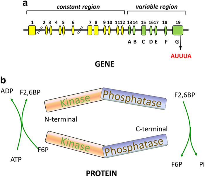
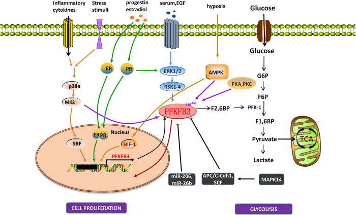
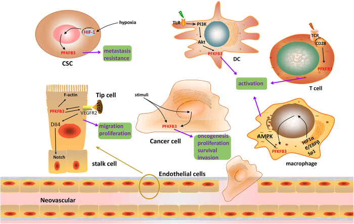
The understanding of 6-phosphofructo-2-kinase/fructose-2,6-biphosphatase 3 (PFK-2/FBPase 3, PFKFB3) has advanced considerably since its initial identification in human macrophages in the mid-1990s. As a vital regulator of glycolysis, accumulating studies have suggested that PFKFB3 is associated with many aspects of cancer, including carcinogenesis, cancer cell proliferation, vessel aggressiveness, drug resistance and tumor microenvironment. In this review, we summarize current knowledge of PFKFB3 regulation by several signal pathways and its function in cancer development in different cell types in cancer tissues. Ubiquitous PFKFB3 has emerged as a potential target for anti-neoplastic therapy.


Conflict of interest statement

The authors declare no conflict of interest.

Figures

Figure 1
Figure 1
General structure of the PFKFB3 gene and protein. (a) The PFKFB3 gene contains at least 19 exons, which can be divided into 2 regions, the constant and variable regions. The variable region contains seven exons named A–G, and variations in the exons in this region leads to six isoforms of PFKFB3. PFKFB3 contains multiple copies of the AUUUA instability element in its 3′UTR. (b) The PFKFB3 protein has two homodimeric subunits. Each subunit of PFKFB3 comprises two functional domains: an N-terminal kinase domain and a C-terminal phosphatase domain. The kinase activity catalyzes the production of F2,6P2 and ADP from F6P and ATP, which highly promote the glycolytic pathway. The phosphatase activity dephosphorylates F2,6P2 to produce F6P and Pi.
Figure 2
Figure 2
Signaling pathways involving PFKFB3. Numerous molecules are associated with PFKFB3 regulation. (1) Progestin, estradiol and hypoxia induce binding of the transcription factors PR, ER and HIF, respectively, to their responsive elements in the PFKFB3 promoter. Inflammatory cytokines and stress stimuli increase PFKFB3 production via the P38/MK2/SRF pathway. Serum and EGF function through the ERK1/2 (extracellular-signal-regulated kinase)/RSK1–4 (ribosomal S6 kinase) pathway, and progestin also regulates glycolysis through this pathway as a secondary mechanism. (2) MiR-206 and miR-26b inhibit PFKFB3 by interacting with 3′UTR of PFKFB3 mRNA. Other negative regulators of PFKFB3, such as ubiquitin ligase APC/C-Cdh1 and SCF, catalyze the degradation of the PFKFB3 protein, which in turn results in decreased glycolysis in cells. (3) PFKFB3 is phosphorylated at Ser461 within the C-terminal region by MK234, AMPK38, PKA and PKC.
Figure 3
Figure 3
Roles of PFKFB3 in different cancer cells. High levels of the PFKFB3 isoenzyme have been proven to promote the oncogenesis, proliferation and survival of cancer cells. Elevated PFKFB3 in CSCs has been estimated to be related to distant metastasis and poor clinical outcome. PFKFB3 is apparently induced by hypoxia in CSCs. Silencing of PFKFB3 impairs vessel sprouting due to defects in both migrating tip and proliferating stalk cells. PFKFB3, compartmentalized with F-actin in lamellipodia, provide ATPs for vascular sprouting, and VEGFR2 induces PFKFB3 and activates Notch signaling. Immune cells shift from OXPHO to glycolysis when activated. The TLR/PI3K/Akt signaling pathway controls this shift in DC cells. In T cells, PFKFB3 is induced downstream by the TCR/CD28 receptor. PFKFB3 expression is increased by the transcription factors HIF1α, C/EBPβ and Sp1 in macrophages, and the PFKFB3 enzyme is phosphorylated by AMPK.

References

    1. Warburg O. On the origin of cancer cells. Science 1956; 123: 309–314. - PubMed
    1. Weber G. Enzymology of cancer cells (first of two parts). N Engl J Med 1977; 296: 486–449. - PubMed
    1. Van Schaftingen E, Jett MF, Hue L, Hers HG. Control of liver 6-phosphofructokinase by fructose 2,6-bisphosphate and other effectors. Proc Natl Acad Sci Biol 1981; 78: 3483–3486. - PMC - PubMed
    1. Pilkis SJ, Claus TH, Kurland IJ, Lange AJ. 6-Phosphofructo-2-kinase/fructose-2,6-bisphosphatase: a metabolic signaling enzyme. Annu Rev Biochem 1995; 64: 799–835. - PubMed
    1. Okar DA, Lange AJ. Fructose-2,6-bisphosphate and control of carbohydrate metabolism in eukaryotes. Biofactors 1999; 10: 1–14. - PubMed

Publication types