. 2017 Oct 27:2:17051.
doi: 10.1038/sigtrans.2017.51. eCollection 2017.

STAT3 mediates C6-ceramide-induced cell death in chronic lymphocytic leukemia

Affiliations

STAT3 mediates C6-ceramide-induced cell death in chronic lymphocytic leukemia

Ushma A Doshi et al. Signal Transduct Target Ther. .

Abstract

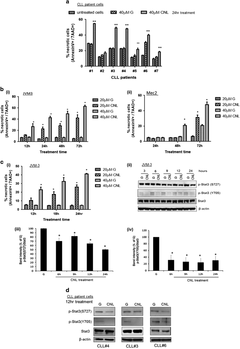
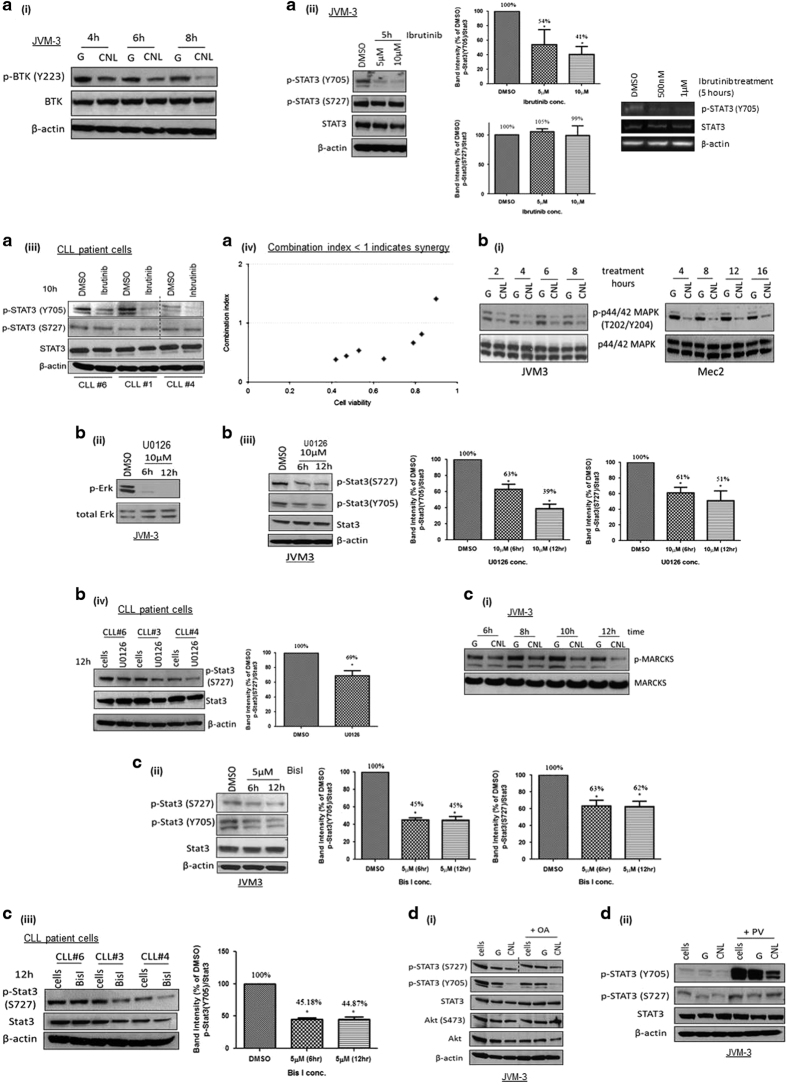
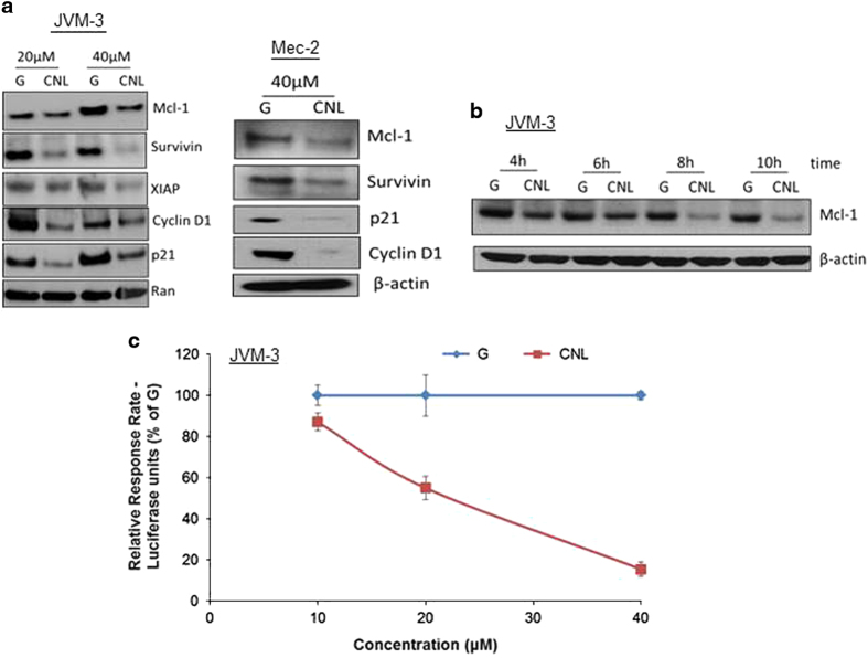
The pathogenesis of chronic lymphocytic leukemia (CLL) is poorly understood and it remains incurable with current therapies. We have previously shown that nanoliposomal C6-ceramide (CNL) is an effective therapy in an in vivo murine model of CLL. However, the key signaling pathways mediating CNL-induced cell death in CLL remains unknown. We hypothesized that CNL targets STAT3, a critical regulator of hematopoietic biology. We observed that CNL treatment reduced phosphorylated STAT3 at both Y705 and S727 residues in CLL cell lines and patient cells. This, in turn, reduced STAT3 transcriptional activity and expression of critical STAT3-dependent survival factors like Mcl-1 and survivin. The effect of CNL on STAT3 was further confirmed ex vivo as shown by reduced STAT3 phosphorylation in xenograft tumors obtained from mice treated with CNL. CNL suppressed STAT3 phosphorylation at Y705 and S727 through reduction in BTK activity and MEK1/2 kinase/PKC activities, respectively. Moreover, a synergistic reduction in CLL cell viability was observed on co-treatment with CNL and the BTK inhibitor, ibrutinib. Expression of an oncogenic form of STAT3 conferred partial resistance to CNL, providing confirmation that STAT3 mediates CNL-induced cell death. Taken together, these findings provide the first body of evidence demonstrating ceramide regulation of STAT3 phosphorylation. These results are also the first to demonstrate an effect of ceramide on BTK, a critical kinase mediating the B-cell receptor signaling in CLL cells and suggest a novel and synergistic combination of CNL and BTK inhibitors for CLL treatment.


Conflict of interest statement

MK—Penn State Research Foundation (PSRF) has licensed ceramide nanoliposomes to Keystone Nano, Inc. PSRF has previously licensed other ceramide nanotechnologies to Keystone Nano, Inc. MK is chief medical officer of Keystone Nano, Inc, State College, PA. The remaining authors declare no competing financial interests.

Figures

Figure 1
Figure 1
STAT3 is a potential therapeutic target in CLL. (a) STAT3 is overexpressed in CLL cell lines and patient cells. (i) JVM-3 cells, Mec-2 cells, PBMCs from two different normal blood donors and PBMCs from four CLL patients were lysed and western blot analysis was performed. The final image was created by grouping different parts of the same film of the same gel as indicated by the black dividing line. (ii) CD19+ B cells were isolated from blood donated by healthy donors and protein levels were compared to JVM-3 cells by western blot analysis. (b) Knockdown of STAT3 induces cell death in CLL cells. JVM-3 cells were transfected with several clones of STAT3 shRNA. (i) Flow cytometric analysis was performed to determine % dead cells 24–96 h after doxycycline induction and (ii) western blot analysis done. Cells nucleofected with TE buffer containing no plasmid were used as a control. An aliquot of 1 μg ml−1 doxycycline was used to induce the expression of STAT3 shRNA 24 h after nucleofection and doxycycline level was maintained during the assay period. The graph represents two independent experiments. Student’s t-test was used for statistical analysis, ***P<0.0001, **P<0.05. The final western blot image was created by grouping different parts of the same film of the same gel as indicated by the black dividing line. (c) STAT3 inhibition reduces viability of CLL cell lines and patient cells. (i) PBMCs from three normal donors and three CLL patients, (ii) JVM-3 cells and (iii) Mec-2 cells were treated with Stattic for 24 h and cell viability was determined by the MTS assay. (ii) Western blotting analysis in JVM-3 confirms the effectiveness of Stattic treatment. The graphs represent results from three independent experiments.
Figure 2
Figure 2
CNL suppresses the phosphorylation of STAT3 at both Y705 and S727 residues. CNL suppresses phosphorylation of STAT3 in (a) CLL patient cells; (b) JVM-3 cells; (c) Mec-2 cells; (d) ex vivo xenograft tumors. Cells were treated with 20 μM and/or 40 μM of ghost nanoliposomes or CNL as indicated in the figure for 24 h (CLL patient cells and JVM-3 cells) or 48 and 72 h (Mec-2 cells). Western blotting analysis was performed. The graphs represent the quantification of western blotting from: (a) 7 CLL patient cells; and (b) three independent experiments. The final western blot image was created by grouping different parts of the same film of the same gel as indicated by the black dividing line. Statistical analysis was performed using Student’s t-test, *P<0.05, **P<0.01. (d) JVM-3 xenograft tumors were obtained from a subcutaneous CLL mouse model in Balb/c Nu/nu mice that were injected with ghost nanoliposomes or CNL (from Ryland et al.). Western blotting was performed for one tumor treated with ghost nanoliposomes and two CNL-treated tumors obtained from two separate mice. (e) CNL does affect cell viability and STAT3 phosphorylation in HEK293 cells. (i) Cell viability of HEK293 cells was determined by MTS assay after 24 h treatment and (ii) western blotting analysis was performed. The results are representative of three independent experiments.
Figure 3
Figure 3
Suppression of STAT3 phosphorylation is specific to STAT3 and C6-ceramide. (a) CNL-induced suppression of phosphorylation is specific to STAT3. JVM-3 cells were treated with 40 μM ghost nanoliposomes or CNL for 24 h and western blotting analysis was done. A positive control of STAT2 phosphorylation at Y690 was also used. The images are representative of three independent experiments. (b) Only C6-ceramide sphingolipid suppresses STAT3 phosphorylation. JVM-3 cells were treated with dihrdro-C6-ceramide nanoliposomes or BSA:sphingosine complex or BSA:S1P complex for 24 h. Western blotting analysis was performed. The images are representative of three independent experiments.
Figure 4
Figure 4
CNL induces necrotic cell death in CLL cells. CNL induces necrotic cell death in (a) CLL patient cells and (b) CLL cell lines JVM-3 and Mec-2. Cells were treated with 20 μM and/or 40 μM ghost nanoliposomes or CNL for indicated time periods. Flow cytometric analysis using Annexin-V and 7AAD staining was performed to determine the effect on cell death. (a) The graph represents the quantification of all seven CLL patient samples. Student’s t-test was used to perform statistical analysis **P<0.01. (b) The graphs represent the quantification of results from three independent experiments. Two-way ANOVA with Tukey’s multiple comparisons test was used to perform statistical analysis *P<0.01. (c) CNL-induced suppression of p-STAT3 precedes induction of cell death (i) JVM-3 cells were treated with ghost nanoliposomes or CNL for indicated time periods and flow cytometric analysis was performed to determine % cell death. The graph is a quantification of three independent experiments. Statistical analysis was done using Student’s t-test *P<0.05 (ii) JVM-3 cells were treated with 40 μM ghost nanoliposomes or CNL for indicated time periods and western blotting was performed. (iii) and (iv) Graphical representation of western blotting. The graph is a quantification of three independent experiments. Statistical analysis was done using Student’s t-test *P<0.05. (d) CNL induces early suppression of p-STAT3 in CLL patient cells. Cells from three CLL patients were treated for 12 h with 40 μM ghost nanoliposomes or CNL and western blotting was done.
Figure 5
Figure 5
CNL suppresses STAT3 phosphorylation via multiple kinases including BTK. (a) (i) CNL suppresses the activity of BTK. JVM-3 cells were treated with 40 μM ghost liposomes or CNL for indicated time periods and western blotting was performed. The blots are a representative of three independent experiments. (ii) and (iii) BTK inhibitors suppress phosphorylation of STAT3 in JVM-3 cells and CLL patient cells. Cells were treated with varying concentrations of ibrutinib for indicated time periods and western blotting was performed. Graphical representation of the western blot is also shown. The blots and graphs are representative of three independent experiments or three CLL patient samples. Student’s t-test was used to perform statistical analysis, *P<0.05. The final western blot image was created by grouping different parts of the same film of the same gel as indicated by the black dividing line. (iv) Synergism analysis of CNL and ibrutinib treatments. JVM-3 cells were treated with single agents and co-treated with different doses of CNL (1–10 μM) and ibrutinib (1–2.5 μM) for 24 h and MTS assay was performed. The cell viability data were analyzed for synergism using Compusyn software. No synergism was observed with ghost nanoliposomes. (b) (i) CNL suppresses the activity of MEK1/2 kinase. JVM-3 and Mec-2 cells were treated with 40 μM ghost liposomes or CNL for indicated time periods and western blotting was performed. The blots are a representative of three independent experiments. (ii) JVM-3 cells were treated with 10 μM U0126 for indicated time periods and p-Erk levels were evaluated to confirm the effectiveness of U0126 as a MEK inhibitor. (iii) and (iv) MEK1/2 inhibitor suppresses phosphorylation of STAT3 in JVM-3 cells and CLL patient cells. Cells were treated with 10 μM U0126 for indicated time periods and western blotting was performed. The blots and graphs are representative of three independent experiments or three CLL patient samples. Student’s t-test was used for statistical analysis, *P<0.05. (c) (i) CNL suppresses the activity of PKC. JVM-3 cells were treated with 40 μM ghost liposomes or CNL for indicated time periods and western blotting was performed. The blots are a representative of three independent experiments. (ii) and (iii) PKC inhibitor suppresses phosphorylation of STAT3 in JVM-3 cells and CLL patient cells. Cells were treated with 5 μM Bis-I for indicated time periods and western blotting was performed. The blots and graphs are representative of three independent experiments or three CLL patient samples. Student’s t-test was used for statistical analysis, *P<0.05. (d) CNL does not activate phosphatases. JVM-3 cells were pretreated for 2 h with: (i) 5 nM OA; (ii) 50 μM PV, followed by 12 h of treatment with 40 μM ghost nanoliposomes or CNL. Both the inhibitors were non-toxic to cells at the specific concentration. The blots are a representative of two independent experiments. The final western blot image was created by grouping different parts of the same film of the same gel as indicated by the black dividing line.
Figure 6
Figure 6
CNL suppresses the transcriptional activity of STAT3. (a) CNL reduces levels of STAT3-regulated genes. JVM-3 cells and Mec-2 cells were treated with 20 μM or 40 μM ghost liposomes or CNL and western blotting was performed. JVM3 cells were treated for 24 h and Mec-2 cells were treated with 48 h. The images are representative of three independent experiments. (b) Reduction of STAT3 phosphorylation precedes reduction of Mcl-1 levels following CNL treatment. JVM-3 cells were treated with 40 μM ghost liposomes or CNL for indicated time periods and western blotting was performed. (c) CNL inhibits expression of luciferase in a STAT3 luciferase reporter assay. JVM-3 cells were transfected with different luciferase constructs. Twelve hours after transfection, cells were treated with ghost nanoliposomes or CNL for 12 h and luciferase assay was performed. The graphs are representative of three independent experiments.
Figure 7
Figure 7
Overexpression of STAT3-C rescues CNL-induced cell death. (a) STAT3-C-expressing cells are resistant to CNL-induced cell death. Lentiviral transduction was performed to express STAT3-C in JVM-3 cells. Seventy-two hours after the last transduction, FACS was performed to obtain a pure population of cells expressing STAT3-C and the treatments were done. An overexpression construct expressing RFP was used as a negative control. Seventy-two hours after the last transduction cycle, cells were treated with ghost liposomes and CNL for 24 h. (i) Expression of STAT3-C was confirmed by western blotting and probing for Flag-tag. (ii) Flow cytometric analysis for Annexin-V and 7AAD was performed to quantitate % necrotic cells. Student’s t-test was used for statistical analysis, *P<0.05.

References

    1. Sutton LA, Rosenquist R. Deciphering the molecular landscape in chronic lymphocytic leukemia: time frame of disease evolution. Haematologica 2015; 100: 7–16. - PMC - PubMed
    1. Zenz T, Mertens D, Kuppers R, Dohner H, Stilgenbauer S. From pathogenesis to treatment of chronic lymphocytic leukaemia. Nat Rev Cancer 2010; 10: 37–50. - PubMed
    1. Tam CS, O'Brien S, Wierda W, Kantarjian H, Wen S, Do KA et al. Long-term results of the fludarabine, cyclophosphamide, and rituximab regimen as initial therapy of chronic lymphocytic leukemia. Blood 2008; 112: 975–980. - PMC - PubMed
    1. Hallek M, Fischer K, Fingerle-Rowson G, Fink AM, Busch R, Mayer J et al. Addition of rituximab to fludarabine and cyclophosphamide in patients with chronic lymphocytic leukaemia: a randomised, open-label, phase 3 trial. Lancet 2010; 376: 1164–1174. - PubMed
    1. Hankins JL, Doshi UA, Haakenson JK, Young MM, Barth BM, Kester M. The therapeutic potential of nanoscale sphingolipid technologies. Handb Exp Pharmacol 2013, 197–210. - PubMed

Publication types

LinkOut - more resources