

Pygopus 2 promotes kidney cancer OS-RC-2 cells proliferation and invasion in vitro and in vivo
- PMID: 29264135
- PMCID: PMC5730714
- DOI: 10.1016/j.ajur.2015.06.009
Pygopus 2 promotes kidney cancer OS-RC-2 cells proliferation and invasion in vitro and in vivo
Abstract
Objective: Human Pygopus 2 (Pygo2) was recently discovered to be a component of the Wnt signaling pathway required for β-catenin/Tcf-mediated transcription. But the role of Pygo2 in malignant cell proliferation and invasion has not yet been determined.
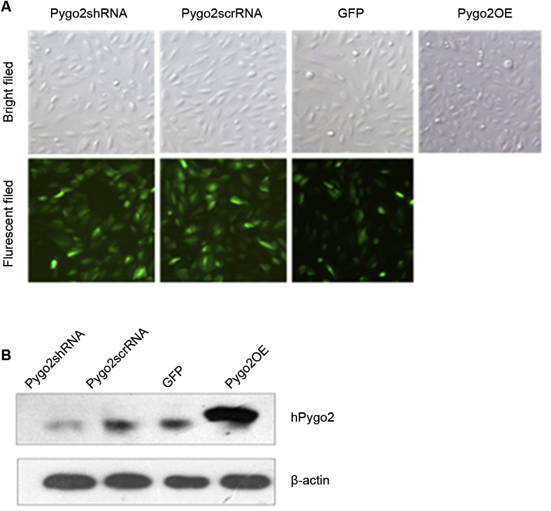
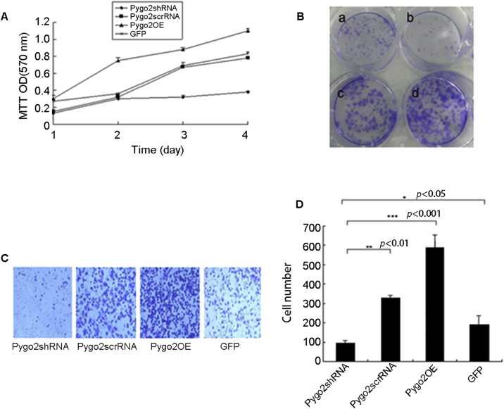
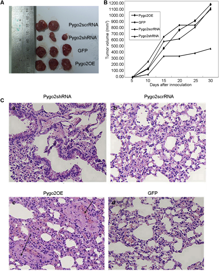
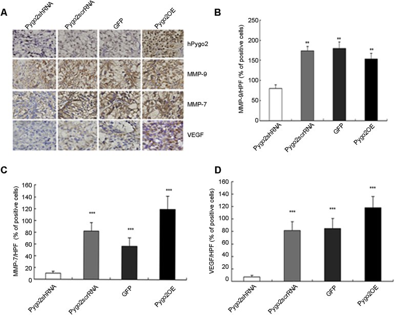
Methods: Lentivirus-mediated small interfering RNA (siRNA) and vector-based overexpression were used to study the function of Pygo2 in OS-RC-2 cells. The resulted cells were subject to Western blotting assay, MTT assay, colony formation and cell invasion assays. Furthermore, renal cell carcinoma (RCC) models were established in BALB/c nude mice inoculated with OS-RC-2 cells. Immunohistochemistry (IHC) staining of matrix metalloproteinase-7 (MMP-7), matrix metalloproteinase-9 (MMP-9) and vascular endothelial growth factor (VEGF) was performed in tumor tissue.
Results: Pygo2 gene was successful knocked down and overexpressed in RCC OS-RC-2 cells by using an shRNA and overexpressing vector, respectively. Overexpression of Pygo2 effectively promoted cell proliferation, colony formation and invasion in vitro. Knockdown of Pygo2 obviously inhibited xenograft tumor growth in nude mice. In addition, overexpression of Pygo2 increased the levels of MMP-7, MMP-9 and VEGF in the xenograft tumors.
Conclusion: Pygo2 has a role in promoting cell proliferation, invasion and metastasis, and may regulate angiogenesis via the Wnt/β-catenin signaling pathway.
Keywords: Matrix metalloproteinase-7; Matrix metalloproteinase-9; Pygo2; Renal cell carcinoma; Vascular endothelial growth factor.
Figures
Similar articles
-
Overexpression of Pygopus-2 is required for canonical Wnt activation in human lung cancer.Oncol Lett. 2014 Jan;7(1):233-238. doi: 10.3892/ol.2013.1691. Epub 2013 Nov 19. Oncol Lett. 2014. PMID: 24348855 Free PMC article.
-
Downregulation of Pygopus 2 inhibits vascular mimicry in glioma U251 cells by suppressing the canonical Wnt signaling pathway.Oncol Lett. 2016 Jan;11(1):678-684. doi: 10.3892/ol.2015.3917. Epub 2015 Nov 13. Oncol Lett. 2016. PMID: 26870266 Free PMC article.
-
The role of Pygopus 2 in rat glioma cell growth.Med Oncol. 2011 Jun;28(2):631-40. doi: 10.1007/s12032-010-9488-1. Epub 2010 Apr 2. Med Oncol. 2011. PMID: 20361361
-
Decreased pygopus 2 expression suppresses glioblastoma U251 cell growth.J Neurooncol. 2010 Oct;100(1):31-41. doi: 10.1007/s11060-010-0144-6. Epub 2010 Mar 5. J Neurooncol. 2010. PMID: 20204459
-
Interference of EFNA4 suppresses cell proliferation, invasion and angiogenesis in hepatocellular carcinoma by downregulating PYGO2.Cancer Biol Ther. 2022 Dec 31;23(1):1-12. doi: 10.1080/15384047.2022.2149039. Cancer Biol Ther. 2022. PMID: 36404439 Free PMC article.
Cited by
-
RAC2 acts as a prognostic biomarker and promotes the progression of clear cell renal cell carcinoma.Int J Oncol. 2019 Sep;55(3):645-656. doi: 10.3892/ijo.2019.4849. Epub 2019 Jul 26. Int J Oncol. 2019. PMID: 31364727 Free PMC article.
-
PYGO2 regulates IL10 and plays immunosuppressive role through ESCC progression.BMC Mol Cell Biol. 2025 Apr 29;26(1):14. doi: 10.1186/s12860-025-00540-0. BMC Mol Cell Biol. 2025. PMID: 40301712 Free PMC article.
-
TRIM2 downregulation in clear cell renal cell carcinoma affects cell proliferation, migration, and invasion and predicts poor patients' survival.Cancer Manag Res. 2018 Nov 20;10:5951-5964. doi: 10.2147/CMAR.S185270. eCollection 2018. Cancer Manag Res. 2018. PMID: 30538545 Free PMC article.
-
Structural basis of the interaction between BCL9-Pygo and LDB-SSBP complexes in assembling the Wnt enhanceosome.Nat Commun. 2023 Jun 22;14(1):3702. doi: 10.1038/s41467-023-39439-9. Nat Commun. 2023. PMID: 37349336 Free PMC article.
-
Histopathological Evaluation of PYGO2 Expression in Esophageal Squamous Cell Carcinoma.Iran J Pathol. 2024 Fall;19(4):415-521. doi: 10.30699/ijp.2024.2024609.3269. Epub 2024 Oct 2. Iran J Pathol. 2024. PMID: 40034933 Free PMC article.
References
-
- Hoffmans R., Städeli R., Basler K. Pygopus and legless provide essential transcriptional coactivator functions to armadillo/beta-catenin. Curr Biol. 2005;15:1207–1211. - PubMed
-
- Kramps T., Peter O., Brunner E., Nellen D., Froesch B., Chatterjee S. Wnt/wingless signaling requires BCL9/legless-mediated recruitment of pygopus to the nuclear β-catenin-TCF complex. Cell. 2002;109:47–60. - PubMed
LinkOut - more resources
Full Text Sources
Other Literature Sources
Miscellaneous
