Review
. 2015 Feb 17;2(2):43-48.
doi: 10.1016/j.asmart.2014.12.003. eCollection 2015 Apr.

One-stage revision anatomic anterior cruciate ligament reconstruction with rectangular tunnel technique

Affiliations
Review

One-stage revision anatomic anterior cruciate ligament reconstruction with rectangular tunnel technique

Konsei Shino et al. Asia Pac J Sports Med Arthrosc Rehabil Technol. .

Abstract

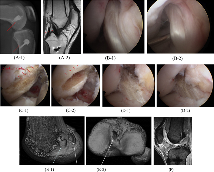
We developed the anatomic rectangular tunnel anterior cruciate ligament reconstruction (ART ACLR) with a bone-patellar tendon-bone graft to mimic fibre arrangement inside the native ACL via tunnels with smaller apertures. With a 10-mm-wide graft, the cross-sectional area of the tunnels of 50 mm2 in ART ACLR is less than that of 79 mm2 in a 10-mm round tunnel one. Because tunnel encroachment would be less of a problem, the ART ACLR technique could be most frequently applied to patients after a failed primary ACLR. In this instructional lecture, the indication and technical considerations for ART ACLR as one-stage revision ACLR are described.

Keywords: anatomic rectangular tunnel technique; bone–patellar tendon–bone graft; one-stage; revision ACL reconstruction.


Figures

Fig. 1
Fig. 1
Intra-articular tunnel apertures of the femoral and the tibial attachment areas for anatomical rectangular tunnel anterior cruciate ligament reconstruction (ART ACLR). (A) Note the tendinous side of the bone plug (black area) located posteriorly–superiorly in the femoral tunnel. (B) The tibial tunnel is almost filled with the tendon (black area).
Fig. 2
Fig. 2
Schema of revision rectangular tunnel ACL reconstruction with BTB graft. The bone plug is fixed to the femur with a 6-mm interference screw (IFS), whereas tibial fixation is achieved with a modified pullout suture technique using the DSP (Double Spike Plate) and a screw. A new anatomic femoral tunnel can be properly placed in most cases without overlapping tunnels despite the previous anterior femoral tunnel (PAFT) leading to a vertical graft. A new tibial tunnel is created to the aperture of the previous vertical tibial tunnel (PVTT) in most cases, whereas the direction of the tunnel is changed.
Fig. 3
Fig. 3
Dilators of four sizes: 13 mm × 6.5 mm, 10 mm × 6.5 mm, 10 mm × 5 mm, and 8 mm × 5 mm.
Fig. 4
Fig. 4
(A-1) Plain lateral radiograph of Case 1. Note the big fixation hardware (unbroken arrows). (A-2) T1-weighted sagittal magnetic resonance (MR) image of Case 1 showing the vertically oriented graft (broken arrow). (B-1) Arthroscopic appearance of the graft in flexion. Note the graft-posterior cruciate ligament (PCL) impingement. (B-2) Arthroscopic appearance of the graft with abnormal looseness in extension. (C-1) Arthroscopic appearance of femoral attachment area behind the resident's ridge (unbroken arrows) via the anteromedial portal, after excising the nonfunctioning graft. (C-2) The new anatomic femoral tunnel aperture just behind the resident's ridge (unbroken arrows). (D-1) Revision anatomic ACL graft in flexion showing no graft-PCL impingement. (D-2) Revision anatomic ACL graft in extension showing no abnormal looseness in extension. (E-1) Three-dimensional (3-D) computed tomography (CT) pictures of Case 1 showing femoral and tibial tunnel apertures after revision ACLR. Note the new anatomic rectangular femoral tunnel aperture (unbroken arrow) and the primary nonanatomic one (broken arrow). (E-2) 3-D CT picture of Case 1 showing the reused tibial tunnel aperture (unbroken arrow). (F) T2-weighted sagittal MR image of Case 1 at 11 months after the revision ACLR. Note that the graft runs properly as the normal ACL (unbroken arrow).
Fig. 5
Fig. 5
(A-1) Three-dimensional (3-D) computed tomography (CT) picture of Case 2 showing improper femoral tunnels (unbroken arrows). (A-2) 3-D CT picture of Case 2 showing too far posterior tibial tunnel aperture (broken arrow). (B-1) Arthroscopic appearance of the primary nonanatomic ACL graft of Case 2. Note its vertical and loose appearance. (B-2) Arthroscopic appearance of the new femoral tunnel in the anatomic attachment area (unbroken arrow), and the primary nonanatomic tunnel aperture (broken arrow) filled with an interference screw. (C-1) 3-D CT picture of Case 2 showing femoral tunnel aperture after the revision ACLR. Note the new anatomic rectangular femoral tunnel aperture (unbroken arrow) and the primary nonanatomic ones (broken arrows). (C-2) 3-D CT picture of Case 2 showing tibial tunnel aperture after the revision ACLR. Note the new tibial tunnel aperture (unbroken arrow) and the primary posterior one (broken arrow).
Fig. 6
Fig. 6
(A-1) Three-dimensional (3-D) computed tomography (CT) picture of Case 3 showing femoral tunnel apertures (unbroken arrows). (A-2) 3-D CT picture of Case 3 showing a single widened tibial tunnel (broken arrow) after the third ACLR. (B-1) 3-D CT picture of Case 3 after the fourth ACLR showing through over the top of the lateral femoral condyle (unbroken arrows). (B-2) CT axial view of the tibial tunnel after the fourth ACLR. Note the bone plug of the quadriceps tendon–bone graft (unbroken arrow) and the cylindrical bone graft behind the bone plug (broken arrow).

Similar articles

Cited by

References

    1. Bach B.R., Jr. Revision anterior cruciate ligament surgery. Arthroscopy. 2003;19:14–29. - PubMed
    1. Siebold R., Schumacher P., Fernandez F. Flat midsubstance of the anterior cruciate ligament with tibial “C”-shaped insertion site. Knee Surg Sports Traumatol Arthosc. 2014 - PMC - PubMed
    1. Noyes F.R., Butler D.L., Grood E.S., Zernicke R.F., Hefzy M.S. Biomechanical analysis of human ligament grafts used in knee-ligament repairs and reconstructions. J Bone Joint Surg. 1984;66-A:344–352. - PubMed
    1. Shino K., Nakata K., Nakamura N., Toritsuka Y., Nakagawa S., Horibe S. Anatomically-oriented ACL Reconstruction with a bone–patellar tendon graft via rectangular socket/tunnels: a snug-fit and impingement-free grafting technique. Arthroscopy. 2005;21:1402.e1–1402.e5. - PubMed
    1. Shino K., Nakata K., Horibe S. Rectangular tunnel double-bundle anterior cruciate ligament reconstruction with bone–patellar tendon–bone graft to mimic natural fiber arrangement. Arthroscopy. 2008;24:1178–1183. - PubMed

LinkOut - more resources