Management of Non-Hodgkin's Lymphoma in Maxillofacial Region with Chemotherapy
- PMID: 29264306
- PMCID: PMC5717915
- DOI: 10.4103/ams.ams_85_17
Management of Non-Hodgkin's Lymphoma in Maxillofacial Region with Chemotherapy
Abstract
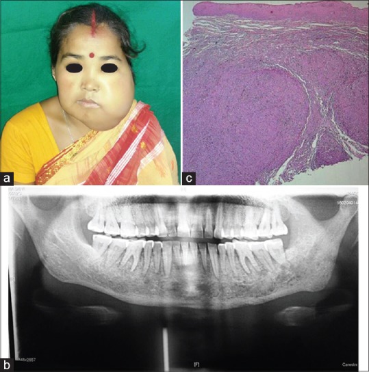
Malignant lymphomas form a heterogeneous group of neoplasms of the lymphoid tissue with different clinical courses, depending on the treatment and prognosis. Lymphoma is a malignant neoplasm of the lymphoid tissue; it is broadly classified into Hodgkin lymphoma (HL) and Non-HL (NHL) depending on the presence or absence of the Reed-Sternberg cells. The main types of lymphomas are (1) HL and (2) NHL. This case report describes about primary NHL involving the mandible. Chemotherapy was advised by the oncologist, and a total of 14 cycles were suggested at the gap of every 2 weeks. The treatment regimen followed was classical Cyclophosphamide hydrodaunorubicin oncovin Prednisolone (CHOP) therapy. NHL can be managed by chemotherapy, radiotherapy, and surgery in various combinations. NHL arising in bone is best treated by chemotherapy and may not require radiotherapy. Survival and prognosis are excellent in localized disease, whereas disseminated disease seems less favorable.
Keywords: CHOP therapy; Chemotherapy; Reed–Sternberg cells; non-Hodgkin's lymphoma; radiotherapy.
Conflict of interest statement
There are no conflicts of interest.
Figures

References
-
- Bunch C, Gatter KC. The lymphomas. In: Weatherall DJ, editor. Oxford Text Book of Medicine. Oxford: Oxford Medical Publications; 1996. pp. 3568–87.
-
- Burke JS. Knowles DM. Neoplastic Hematopathology. 2nd ed. Philadelphia: Lippincott Williams and Willkins; 2001. Waldeyer's ring, sinonasal region, salivary gland, thyroid gland, Central nervous system, and other extranodal lymphomas and lymphoid hyperplasias; pp. 1351–90.
-
- Ferry JA, Harris NL. Lymphomas and lymphoid hyperplasia in head and neck sites. In: Pilch BZ, editor. Head and Neck Surgical Pathology. 1st ed. Philadelphia: Lippincott Williams and Wilkins; 2001. pp. 476–533.
-
- Richards A, Costelloe MA, Eveson JW, Scully C, Irvine GH, Rooney N. Oral mucosal non-Hodgkin's lymphoma – A dangerous mimic. Oral Oncol. 2000;36:556–8. - PubMed
Publication types
LinkOut - more resources
Full Text Sources
Other Literature Sources
Research Materials
Miscellaneous