. 2018 Jul;23(4):595-607.
doi: 10.1007/s12192-017-0868-8. Epub 2017 Dec 20.

Oxidative stress triggers aggregation of GFP-tagged Hsp31p, the budding yeast environmental stress response chaperone, and glyoxalase III

Affiliations

Oxidative stress triggers aggregation of GFP-tagged Hsp31p, the budding yeast environmental stress response chaperone, and glyoxalase III

Urszula Natkańska et al. Cell Stress Chaperones. 2018 Jul.

Abstract

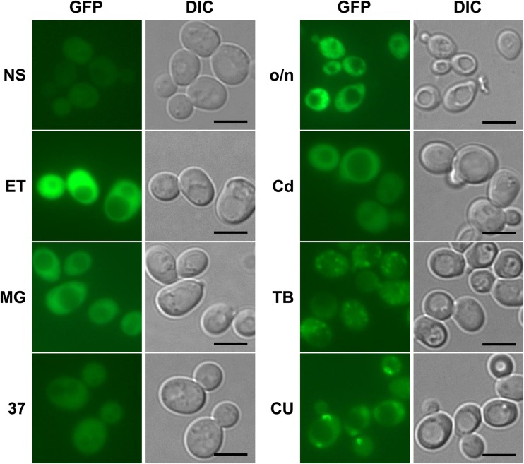
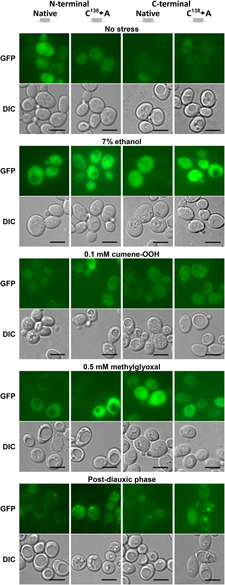
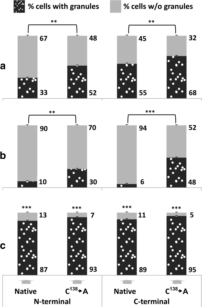
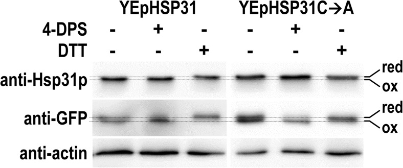
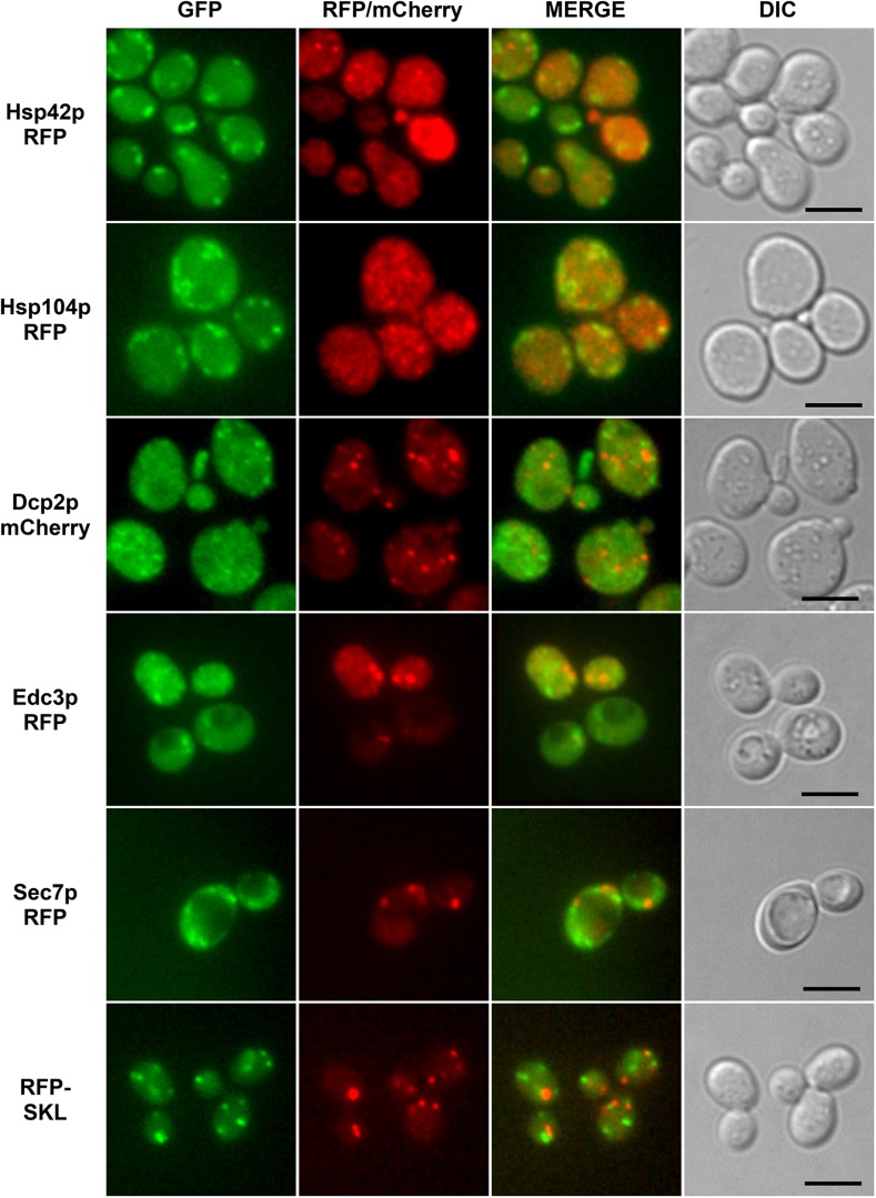
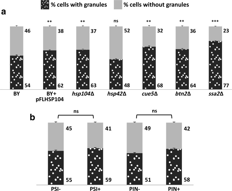
The Saccharomyces cerevisiae Hsp31p protein belongs to the ubiquitous DJ-1/ThiJ/PfpI family. The most prominent member of this family is human DJ-1; defects of this protein are associated with Parkinson's disease pathogenesis. Numerous recent findings reported by our group and others have revealed the importance of Hsp31p for survival in the post-diauxic phase of cell growth and under diverse environmental stresses. Hsp31p was shown to possess glutathione-independent glyoxalase III activity and to function as a protein chaperone, suggesting that it has multiple cellular roles. Our previous work also revealed that HSP31 gene expression was controlled by multiple stress-related transcription factors, which mediated HSP31 promoter responses to oxidative, osmotic, and thermal stresses, toxic products of glycolysis, and the diauxic shift. Nevertheless, the exact role of Hsp31p within budding yeast cells remains elusive. Here, we aimed to obtain insights into the function of Hsp31p based on its intracellular localization. We have demonstrated that the Hsp31p-GFP fusion protein is localized to the cytosol under most environmental conditions and that it becomes particulate in response to oxidative stress. However, the particles do not colocalize with other granular subcellular structures present in budding yeast cells. The observed particulate localization does not seem to be important for Hsp31p functionality. Instead, it is likely the result of oxidative damage, as the particle abundance increases when Hsp31p is nonfunctional, when the cellular oxidative stress response is affected, or when cellular maintenance systems that optimize the state of the proteome are compromised.

Keywords: Environmental stresses; Protein aggregates; Protein stability; Saccharomyces cerevisiae.


Figures

Fig. 1
Fig. 1
Intracellular localization of C-terminally GFP-tagged Hsp31p expressed from the genomic locus under standard growth conditions and after exposure to various stresses. S. cerevisiae cells of the YDR533C clone from the YeastGFP collection (Huh et al. 2003) were grown at 28 °C in SC medium either overnight to reach the post-diauxic growth phase (o/n) or to mid-exponential phase (1–2 × 107 cells × ml−1) (NS), followed by 2 h of exposure to 7% ethanol (ET), 0.5 mM methylglyoxal (MG), 1.25 μM cadmium chloride (Cd), 0.2 mM tert-butyl-OOH (TB), or 0.1 mM cumene-OOH (CU). To apply heat-stress conditions, yeast cells pregrown to mid-exponential phase at 23 °C were transferred to a 37 °C water bath and incubated for 30 min with shaking. Following incubation under stress conditions, the cells were spun down and resuspended in PBS, and GFP fluorescence was examined on an Axio Imager.M2 fluorescence microscope using a 100× objective and 38HE-GFP filter set (Zeiss). Nomarski optics were used for bright-field cell visualization (DIC). Scale bar: 5 μm
Fig. 2
Fig. 2
Neither the presence of the Cys138 amino acid residue nor the placement of the GFP tag is critical for the intracellular distribution of GFP-tagged Hsp31p. Wild-type BY4741 cells were transformed with single-copy YCp50-derivative plasmids bearing constructs encoding either native Hsp31p or Hsp31p lacking the conserved cysteine residue (C138➔A), which were each tagged with GFP at the N- or C-terminus (see the “Materials and methods” section). Transformed cells were grown, exposed to stressors, and visualized by fluorescence microscopy as described in Fig. 1 legend. Scale bar: 5 μm
Fig. 3
Fig. 3
Quantitative analysis of the intracellular distribution of GFP-tagged Hsp31p in wild-type and yap1Δ strains exposed to oxidative stress and in wild-type post-diauxic cells. Wild-type (a, b) and yap1Δ (c) cells were transformed with the same plasmids described in Fig. 2 legend. Wild-type (a) and yap1Δ (c) transformant cells were grown to mid-exponential phase (1–2 × 107 cells × ml−1) in SC medium and exposed to 0.1 mM cumene-OOH for 2 h. Wild-type transformant cells (b) were grown overnight in SC medium. Then, the cells were visualized by fluorescence microscopy as described in Fig. 1 legend. Cells of the transformant strains were scored for the presence of green fluorescent particles. Each data point is the average of three biological replicates, with 200 cells counted per replicate; the standard deviations are represented by error bars. Statistical significance was calculated with Student’s t test: *** p < 0.001, ** 0.001 < p < 0.05. Panels (a) and (b) show the significance of the differences between the native proteins and proteins lacking the Cys138 residue. The significance shown in panel (c) refers to the differences between the data obtained for the yap1Δ deletion strain and the wild type (a) transformed with the respective plasmids
Fig. 4
Fig. 4
The conserved Cys138 amino acid residue of Hsp31p is susceptible to oxidation in cells exposed to oxidative stress. hsp31Δ strains transformed with multi-copy plasmids expressing either native Hsp31p (YEpHSP31) or Hsp31p without the Cys138 residue (YEpHSP31C➔A) and with the plasmid expressing the reference redox-sensitive rxYFP protein were grown to exponential phase, exposed to oxidative stress and then treated with either oxidizing (4-DPS) or reducing (DTT) agents or left untreated. The electrophoretic mobility of Hsp31p and rxYFP was analyzed by non-denaturing SDS polyacrylamide gel electrophoresis, followed by Western blotting and immunodetection of proteins with the anti-Hsp31p-HRP, anti-GFP-HRP primary conjugated antibodies, or anti-actin antibodies as the loading control (see the “Materials and methods” section for more details). The “red” and “ox” terms denote the reduced and oxidized forms of the proteins, respectively
Fig. 5
Fig. 5
GFP-tagged Hsp31p does not colocalize with marker proteins of particulate structures found in budding yeast cells exposed to oxidative stress. Wild-type BY4741 cells were transformed with a single-copy YCp50-derivative plasmid expressing C-terminally GFP-tagged Hsp31p and with plasmids expressing markers of stress granules (Dcp2p-mCherry), P-bodies (Edc3p-RFP), Golgi apparatus (Sec7p-RFP) or peroxisomes (mRFP-SKL). Additionally, the colocalization of Hsp31p-GFP and Hsp42p-RFP or Hsp104p-RFP, all of which form particles in cells exposed to oxidative stress, was analyzed in the same manner. See Table 2 for the list of plasmids used in this experiment. The transformant strains were grown to mid-exponential phase in SC medium and then exposed to 0.1 mM cumene-OOH for 2 h. GFP, RFP, and mCherry fluorescence was examined on an Axio Imager.M2 fluorescence microscope using a 100× objective and 38HE-GFP or 20HE-rhodamine filter sets (Zeiss)
Fig. 6
Fig. 6
The assembly of Hsp31p-GFP particles is inhibited by cellular chaperone proteins but is not influenced by prion formation. (a) Wild-type strain (BY), the same strain overproducing the Hsp104p chaperone (BY + pFLHSP104) and the indicated deletion strains (hsp104Δ, hsp42Δ, cue5Δ, btn2Δ, and ssa2Δ) or (b) the strains containing the [PSI+] or [PIN+] prions together with their prionless isogenic parental strains (see Table 1 for the detailed strain descriptions) were transformed with a single-copy YCp-derivative plasmid expressing C-terminally tagged Hsp31p. The transformant strains were grown to mid-exponential phase in SC medium, exposed to 0.1 mM cumene-OOH for 2 h, visualized by fluorescence microscopy and scored for the presence of green fluorescence particles as described in Fig. 3 legend. Each data point is the average of three biological replicates, with 200 cells counted per replicate; the standard deviations are represented by error bars. Statistical significance was calculated with Student’s t test: *** p < 0.001, ** 0.001 < p < 0.05, ns p > 0.05. Significance in panel (a) refers to the first (BY) data point

Similar articles

Cited by

References

    1. Amm I, Norell D, Wolf DH. Absence of the yeast Hsp31 chaperones of the DJ-1 superfamily perturbs cytoplasmic protein quality control in late growth phase. PLoS One. 2015;10(10):e0140363. doi: 10.1371/journal.pone.0140363. - DOI - PMC - PubMed
    1. Antonenkov VD, Grunau S, Ohlmeier S, Hiltunen JK. Peroxisomes are oxidative organelles. Antioxid Redox Signal. 2010;13(4):525–537. doi: 10.1089/ars.2009.2996. - DOI - PubMed
    1. Aslam K, Hazbun TR. Hsp31, a member of the DJ-1 superfamily, is a multitasking stress responder with chaperone activity. Prion. 2016;10(2):103–111. doi: 10.1080/19336896.2016.1141858. - DOI - PMC - PubMed
    1. Aslam K, Tsai C-J, Hazbun TR. The small heat shock protein Hsp31 cooperates with Hsp104 to modulate Sup35 prion aggregation. Prion. 2016;10(6):444–465. doi: 10.1080/19336896.2016.1234574. - DOI - PMC - PubMed
    1. Bankapalli K, Saladi S, Awadia SS, et al. Robust glyoxalase activity of Hsp31, a ThiJ/DJ-1/PfpI family member protein, is critical for oxidative stress resistance in Saccharomyces cerevisiae. J Biol Chem. 2015;290(44):26491–26507. doi: 10.1074/jbc.M115.673624. - DOI - PMC - PubMed

Publication types

MeSH terms

LinkOut - more resources