. 2018 Jul;55(7):6169-6181.
doi: 10.1007/s12035-017-0834-6. Epub 2017 Dec 20.

A Novel Ultrasensitive In Situ Hybridization Approach to Detect Short Sequences and Splice Variants with Cellular Resolution

Affiliations

A Novel Ultrasensitive In Situ Hybridization Approach to Detect Short Sequences and Splice Variants with Cellular Resolution

Larissa Erben et al. Mol Neurobiol. 2018 Jul.

Abstract

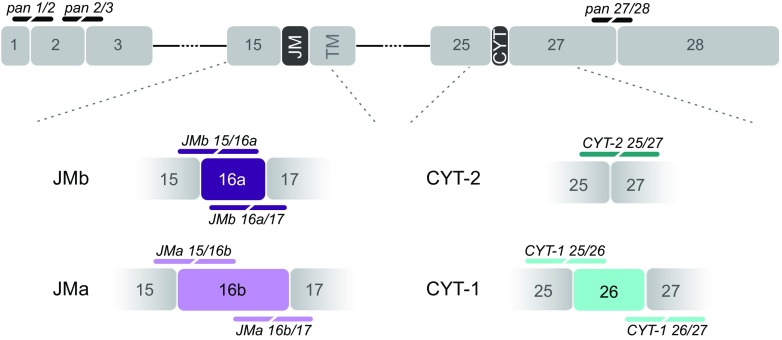
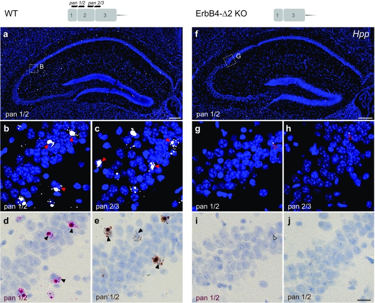
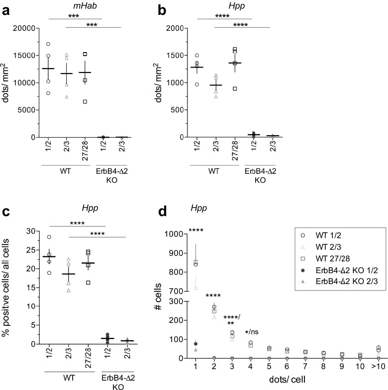
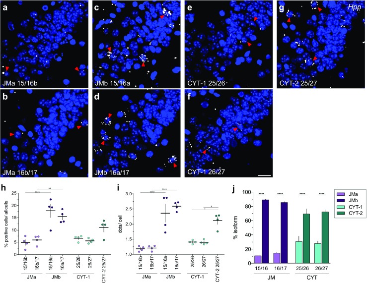
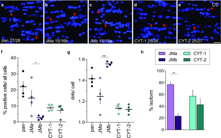
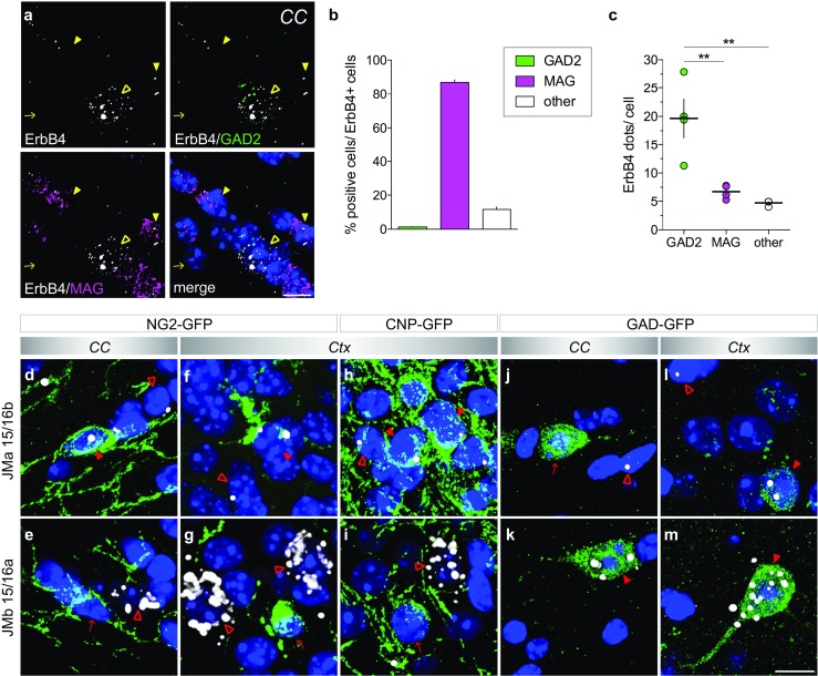
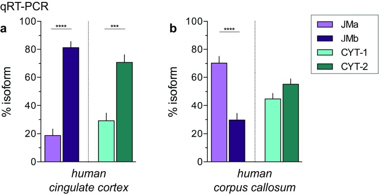
Investigating the expression of RNAs that differ by short or single nucleotide sequences at a single-cell level in tissue has been limited by the sensitivity and specificity of in situ hybridization (ISH) techniques. Detection of short isoform-specific sequences requires RNA isolation for PCR analysis-an approach that loses the regional and cell-type-specific distribution of isoforms. Having the capability to distinguish the differential expression of RNA variants in tissue is critical because alterations in mRNA splicing and editing, as well as coding single nucleotide polymorphisms, have been associated with numerous cancers, neurological and psychiatric disorders. Here we introduce a novel highly sensitive single-probe colorimetric/fluorescent ISH approach that targets short exon/exon RNA splice junctions using single-pair oligonucleotide probes (~ 50 bp). We use this approach to investigate, with single-cell resolution, the expression of four transcripts encoding the neuregulin (NRG) receptor ErbB4 that differ by alternative splicing of exons encoding two juxtamembrane (JMa/JMb) and two cytoplasmic (CYT-1/CYT-2) domains that alter receptor stability and signaling modes, respectively. By comparing ErbB4 hybridization on sections from wild-type and ErbB4 knockout mice (missing exon 2), we initially demonstrate that single-pair probes provide the sensitivity and specificity to visualize and quantify the differential expression of ErbB4 isoforms. Using cell-type-specific GFP reporter mice, we go on to demonstrate that expression of ErbB4 isoforms differs between neurons and oligodendrocytes, and that this differential expression of ErbB4 isoforms is evolutionarily conserved to humans. This single-pair probe ISH approach, known as BaseScope, could serve as an invaluable diagnostic tool to detect alternative spliced isoforms, and potentially single base polymorphisms, associated with disease.

Keywords: Alternative splicing; BaseScope; ErbB4; Neuregulin; Oligodendrocytes; RNA expression; Schizophrenia; Transcriptome.


Conflict of interest statement

L. E. and A.B. declare no competing financial interests. MX.H., A.L. and E.P. are employed by Advanced Cell Diagnostics.

Figures

Fig. 1
Fig. 1
Scheme summarizing ErbB4 isoforms and single-pair probe design. ErbB4 isoforms are generated by alternative splicing of exons encoding the extracellular juxtamembrane domain, resulting in mutually exclusive JMa (exon 16b, light purple, 75 bp) or JMb (exon 16a, dark purple, 45 bp) isoforms, and by inclusion or exclusion of exon 26 encoding a region of the cytoplasmic domain giving rise to CYT-1 (light cyan, 48 bp) and CYT-2 (dark cyan) isoforms, respectively. Single-pair probes targeting all ErbB4 isoforms (pan 1/2, pan 2/3 and pan 27/28) are illustrated in black, whereas isoform-specific single-pair probes targeting splice junctions are color-matched with their respective isoforms. JM: juxtamembrane region; TM: transmembrane domain; CYT: cytoplasmic region
Fig. 2
Fig. 2
Single-pair probes targeting unique exon junctions are specific and sensitive. The specificity and sensitivity of single-pair probes targeting exon-exon boundaries were determined by hybridizing sections from WT (a-e) and ErbB4-Δ2 KO mice (f-j). Probes targeting the exon 1/2 (pan1/2; a,b,d,e) or exon 2/3 (pan 2/3; c) junctions—common to all ErbB4 isoforms—labeled scattered cells in the WT hippocampus (arrowheads). (f-j) By contrast, neither probe generated signals in sections from ErbB4-Δ2 KO mice (background signal marked by open arrowheads). (b, g) Magnified insets in panels (a) and (f) are from area CA2. Signal can be detected by alkaline phosphatase and FastRED visible both in fluorescence (a-c, f-j) and bright field microscopy (d, i) or horseradish peroxidase and diaminobenzidine (e, j). Scale bars: a, f 200 μm; j 20 μm
Fig. 3
Fig. 3
Detection levels for independent probes targeting distinct exon junctions are similar and differ markedly from background in ErbB4-Δ2 KOs. In situ hybridization signals of single-pair probes pan1/2 and pan 2/3 are significantly lower in sections from ErbB4-Δ2 KO mice compared to WT mice in the (a) medial habenula (mHab) and (b) hippocampus (Hpp) (n = 4; one-way ANOVA, see Table S1) and did not differ among pan 1/2, pan 2/3 and pan 27/28 probes in sections from WT mice. (c) Percentage of positive cells relative to all cells in WT hippocampus (CA1–CA3). (d) Histogram distribution of dots/ positive cell detected with single-pair panErbB4 probes in hippocampal CA1–CA3 on sections from WT and ErbB4-Δ2 KO mice. Significance shown for comparisons between WT 1/2 vs. KO 1/2 and WT 2/3 vs. KO 2/3, respectively (n = 4; two-way ANOVA, see Table S4). Adjusted p values according to Tukey’s multiple comparison test: *p < 0.05, **p < 0.01, ***p < 0.001, ****p < 0.0001
Fig. 4
Fig. 4
JMb- and CYT-2-containing transcripts are the major ErbB4 isoforms expressed in adult hippocampus. (ag) Hybridization of ErbB4 isoform-specific single-pair probes in hippocampal CA2 area of WT mice. Arrowheads indicate examples of positive cells. (h, i) Percentages of positive cells/total cells and average dots/cell in hippocampal CA1–CA3 areas were quantified for each isoform-specific probe using CellProfiler. Results derived with probes targeting the same isoform were not significantly different (n = 4; one-way ANOVA, see Table S2). (j) Relative abundance of JMa/JMb (purple) and CYT-1/CYT-2 (cyan) isoforms in the hippocampus (n = 4; one-way ANOVA, see Table S2). Adjusted p values according to Tukey’s multiple comparison test: **p < 0.01, ****p < 0.0001. Scale bar: 20 μm
Fig. 5
Fig. 5
Pattern of ErbB4 JMa and CYT-1 isoform expression in the corpus callosum differ markedly from other brain areas. (ae) Representative in situ hybridization images hybridized with pan and isoform-specific probes in the corpus callosum (CC). Arrowheads indicate representative positive cells. The (f) percentage of positive cells, (g) average number of dots/positive cell and (h) relative expression levels of ErbB4 JMa/JMb and CYT-1/CYT-2 isoforms were quantified using CellProfiler (n = 4; one-way ANOVA, *p < 0.05, **p < 0.01, ***p < 0.001, see Table S3). Scale bar: 20 μm
Fig. 6
Fig. 6
Oligodendrocytes and GABAergic neurons in the corpus callosum express different ErbB4 juxtamembrane isoforms. (a) Multiplex fluorescent in situ hybridization shows that ErbB4 (white) is expressed in both GAD2-positive GABAergic neurons (green; open yellow arrowheads) and MAG-positive oligodendrocytes (magenta; yellow arrowheads) in the corpus callosum (arrow ErbB4-negative cell). Note that dots are smaller compared to single-pair probe ISH, as signals are not enzymatically amplified. (b, c) Quantification of data shown in A (n = 4). (b) The majority of ErbB4+ cells in the corpus callosum co-expresses the oligodendrocytes marker MAG (86.95 ± 1.54%), whereas a small fraction is positive for the GABAergic marker GAD2 (1.40 ± 0.23%); 11.65 ± 1.48% of ErbB4+ cells were not labeled with either marker. (c) However, GABAergic neurons express higher levels of ErbB4 per cell than oligodendrocytes (19.65 ± 3.39 dots/cell vs. 6.73 ± 0.61 dots/cell, p = 0.0034; GAD2 vs. other 4.72 ± 0.23 dots/cell, p = 0.0013 n = 4; MAG vs. other p = 0.7614; F(2,9) = 16.53, p = 0.001; one-way ANOVA; Tukey’s multiple comparisons test: **p < 0.01). (dm) Isoform-specific in situ hybridization using probes JMa 15/16b (d, f, h, j, l) and JMb 15/16a (e, g, i, k, m) was combined with post hoc immunohistochemistry for GFP (green) on sections from NG2-GFP (dg), CNP-GFP (h, i) and GAD-GFP (jm) transgenic mice. JM isoforms (white) were detected on GFP+ cells (red arrowheads), as well as on GFP negative cells (open red arrowheads) in the corpus callosum (CC) and the cortex (Ctx). Arrows depict GFP+ cells negative for JM probes. Note that the detection of JM isoforms in the corpus callosum of CNP-GFP mice was not possible because of the high density of GFP+ myelin sheaths [50]. Scale bar: 10 μm
Fig. 7
Fig. 7
Distinct patterns of ErbB4 JM and CYT isoforms in the gray and white matter are conserved between humans and mice. Relative abundance of JMa/JMb (purple) and CYT-1/CYT-2 (cyan) isoforms in the adult human cingulate cortex (a) and corpus callosum (b) was determined by TaqMan qRT-PCR (n = 4; one-way ANOVA, see Table S5). Adjusted p values according to Tukey’s multiple comparison test: ***p < 0.001, ****p < 0.0001

References

    1. Wang ET, Sandberg R, Luo S, Khrebtukova I, Zhang L, Mayr C, Kingsmore SF, Schroth GP, Burge CB. Alternative isoform regulation in human tissue transcriptomes. Nature. 2008;456(7221):470–476. doi: 10.1038/nature07509. - DOI - PMC - PubMed
    1. Iijima T, Hidaka C, Iijima Y. Spatio-temporal regulations and functions of neuronal alternative RNA splicing in developing and adult brains. Neurosci Res. 2016;109:1–8. doi: 10.1016/j.neures.2016.01.010. - DOI - PubMed
    1. Kang HJ, Kawasawa YI, Cheng F, Zhu Y, Xu X, Li M, Sousa AM, Pletikos M, Meyer KA, Sedmak G, Guennel T, Shin Y, Johnson MB, Krsnik Z, Mayer S, Fertuzinhos S, Umlauf S, Lisgo SN, Vortmeyer A, Weinberger DR, Mane S, Hyde TM, Huttner A, Reimers M, Kleinman JE, Sestan N. Spatio-temporal transcriptome of the human brain. Nature. 2011;478(7370):483–489. doi: 10.1038/nature10523. - DOI - PMC - PubMed
    1. Zhang Y, Chen K, Sloan SA, Bennett ML, Scholze AR, O'Keeffe S, Phatnani HP, Guarnieri P, Caneda C, Ruderisch N, Deng S, Liddelow SA, Zhang C, Daneman R, Maniatis T, Barres BA, Wu JQ. An RNA-sequencing transcriptome and splicing database of glia, neurons, and vascular cells of the cerebral cortex. J Neurosci. 2014;34(36):11929–11947. doi: 10.1523/JNEUROSCI.1860-14.2014. - DOI - PMC - PubMed
    1. Mei L, Xiong WC. Neuregulin 1 in neural development, synaptic plasticity and schizophrenia. Nat Rev Neurosci. 2008;9(6):437–452. doi: 10.1038/nrn2392. - DOI - PMC - PubMed

MeSH terms