

Bone marrow lesions in osteoarthritis: What lies beneath
- PMID: 29266428
- PMCID: PMC8607515
- DOI: 10.1002/jor.23844
Bone marrow lesions in osteoarthritis: What lies beneath
Abstract
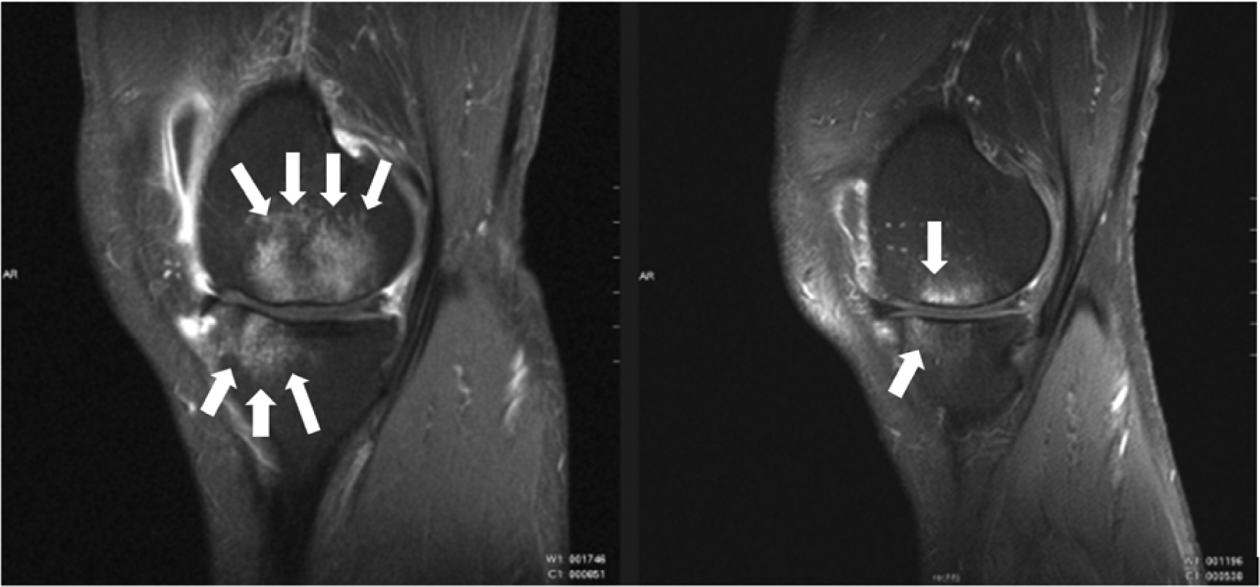
Osteoarthritis (OA) is the most common joint disease in the United States, affecting more than 30 million people, and is characterized by cartilage degeneration in articulating joints. OA can be viewed as a group of overlapping disorders, which result in functional joint failure. However, the precise cellular and molecular events within which lead to these clinically observable changes are neither well understood nor easily measurable. It is now clear that multiple factors, in multiple joint tissues, contribute to degeneration. Changes in subchondral bone are recognized as a hallmark of OA, but are normally associated with late-stage disease when degeneration is well established. However, early changes such as Bone Marrow Lesions (BMLs) in OA are a relatively recent discovery. BMLs are patterns from magnetic resonance images (MRI) that have been linked with pain and cartilage degeneration. Their potential utility in predicting progression, or as a target for therapy, is not yet fully understood. Here, we will review the current state-of-the-art in this field under three broad headings: (i) BMLs in symptomatic OA: malalignment, joint pain, and disease progression; (ii) biological considerations for bone-cartilage crosstalk in joint disease; and (iii) mechanical factors that may underlie BMLs and drive their communication with other joint tissues. Thus, this review will provide insights on this topic from a clinical, biological, and mechanical perspective. © 2017 Orthopaedic Research Society. Published by Wiley Periodicals, Inc. J Orthop Res 36:1818-1825, 2018.
Keywords: Pain; TGF-β; bone-cartilage crosstalk; microdamage; subchondral.
© 2017 Orthopaedic Research Society. Published by Wiley Periodicals, Inc.
Figures


References
-
- Taljanovic MS, Graham AR, Benjamin JB, Gmitro AF, Krupinski EA, Schwartz SA, Hunter TB, and Resnick DL (2008). Bone marrow edema pattern in advanced hip osteoarthritis: quantitative assessment with magnetic resonance imaging and correlation with clinical examination, radiographic findings, and histopathology. Skeletal Radiol 37, 423–431. - PubMed
-
- Felson DT, McLaughlin S, Goggins J, LaValley MP, Gale ME, Totterman S, Li W, Hill C, and Gale D (2003). Bone marrow edema and its relation to progression of knee osteoarthritis. Ann Intern Med 139, 330–336. - PubMed
-
- Felson DT, Chaisson CE, Hill CL, Totterman SM, Gale ME, Skinner KM, Kazis L, and Gale DR (2001). The association of bone marrow lesions with pain in knee osteoarthritis. Ann Intern Med 134, 541–549. - PubMed
Publication types
MeSH terms
Substances
Grants and funding
LinkOut - more resources
Full Text Sources
Other Literature Sources
Medical
