Case Reports
. 2018 Mar;39(3):383-388.
doi: 10.1002/humu.23385. Epub 2018 Jan 13.

Nemaline myopathy and distal arthrogryposis associated with an autosomal recessive TNNT3 splice variant

Affiliations
Case Reports

Nemaline myopathy and distal arthrogryposis associated with an autosomal recessive TNNT3 splice variant

Sarah A Sandaradura et al. Hum Mutat. 2018 Mar.

Abstract

A male neonate presented with severe weakness, hypotonia, contractures and congenital scoliosis. Skeletal muscle specimens showed marked atrophy and degeneration of fast fibers with striking nemaline rods and hypertrophy of slow fibers that were ultrastructurally normal. A neuromuscular gene panel identified a homozygous essential splice variant in TNNT3 (chr11:1956150G > A, NM_006757.3:c.681+1G > A). TNNT3 encodes skeletal troponin-Tfast and is associated with autosomal dominant distal arthrogryposis. TNNT3 has not previously been associated with nemaline myopathy (NM), a rare congenital myopathy linked to defects in proteins associated with thin filament structure and regulation. cDNA studies confirmed pathogenic consequences of the splice variant, eliciting exon-skipping and intron retention events leading to a frameshift. Western blot showed deficiency of troponin-Tfast protein with secondary loss of troponin-Ifast . We establish a homozygous splice variant in TNNT3 as the likely cause of severe congenital NM with distal arthrogryposis, characterized by specific involvement of Type-2 fibers and deficiency of troponin-Tfast .

Keywords: TNNT3; genetics; nemaline myopathy; neuromuscular disease; troponin T-fast.


Figures

Figure 1
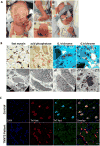
Figure 1. Clinical and pathological features in recessive TNNT3 myopathy with deficiency of troponin Tfast protein demonstrated by immunohistochemistry
A) Clinical photographs at 2 weeks (i), 1 day (ii and iii) and 9 weeks (iv) of age. The proband showed severe weakness of facial and limb muscles, congenital contractures, overlapping digits (i, iii, iv) and congenital vertical talus (ii). B) Immunohistochemistry (IHC) with fast myosin (i) shows atrophic fast fibres, and hypertrophic slow fibres. Average diameter of hypertrophied slow fibres was 40 μm; usual mean diameter of type 1 fibres at 3 months of age on frozen section is 12-13 μm. Acid phosphatase (ii) shows evidence for degeneration of small atrophic fast fibres. Gomori trichrome (iii-iv) and electron microscopy (v-viii) identifies numerous nemaline bodies within atrophic small fibres. v) Lower power electron microscopy image shows one fibre of normal diameter with normal sarcomeric register (top right of field), immune cells (arrows) and numerous atrophied small fibres (asterisks) bearing small electron dense bodies. vi-vii) Electron microscopy images of increasing magnification show numerous nemaline bodies within atrophied fibres. C) IHC of skeletal muscle cryosections from the TNNT3-myopathy proband, reveals atrophied Type 2 fibres positive for fast-myosin (red) that show deficiency/markedly reduced levels of TnTfast (green; data using the Sigma antibody shown). A field with oblique atrophied fast fibres is shown in C, to highlight the absence of detectable labelling for TnTfast (green). Scale bar 25 microns.
Figure 2
Figure 2. mRNA studies confirm pathogenic outcomes arising from the TNNT3 essential splice donor variant
A) i)RT-PCR of muscle cDNA from the TNNT3 proband. Lane 1: Exon 11 forward primer and intron 14 reverse primer show higher levels of intron 14 retention in the patient, relative to an age-matched disease control (male, 4 months, quadricep, mitochondrial myopathy). Lane 2: Exon 11 forward primer and Exon 16 reverse primer amplified a smaller band in the proband, relative to the control. Sanger sequencing showed skipping of exon 14, and roughly equal levels of transcripts splicing from exon 13 – exon 15a – exon 16, or exon 13 – exon 15 – exon 16. RNA sequencing failed for the TNNT3 sample. Thus, relative levels of intron 14 retention versus exon 14 skipping were unable to be determined. ii-iii) Repeat of RT-PCR described in (i), using 30 and 35 cycles to control for saturation, and a second disease control (male, 4 months, quadricep, myotubular myopathy). B) Sashimi plots showing RNA sequencing coverage across TNNT3 exons 12 – 16 (www.gtexportal.org) in controls, showing alternative splicing of either exon 15a or 15. The functional significance of inclusion of either exon is not understood. Sashimi plots also demonstrate low levels of intron 14 retention in controls. Control 1: female, age 33 yrs, deceased tissue donor, left gastrocnemius, cause of death cardiovascular. Disease Control 2: male, age 18 yrs, right quadricep, mild Becker muscular dystrophy (due to DMD splice variant). C) Left: Western blot normalizing for fast myosin content showed marked reduction to near deficiency of TnTfast, and secondary reduction in TnIfast, in the TNNT3-myopathy proband (P1, quadriceps femoris, corrected age 1 month) relative to two age-matched controls (C1: male, vastus lateralis, 4 months; C2: male, vastus lateralis, 4 months). No evidence for a truncated TnTfast protein, or higher molecular weight TnTfast protein, was observed. Right: Specificity of TnTfast and TnIfast antibodies for fast troponin isoforms was validated via western blot of single fibres (fast Type 2A or slow Type 1) dissected from human quadriceps. D) Schematic of TNNT3 exon structure and consequences of the TNNT3 splicing variant on the encoded troponin-Tfast protein. Upper: Exon structure of the TNNT3 gene based on collective information from (Stefancsik et al., 2003), https://genome.ucsc.edu/, www.gtexportal.org/. Alternately spliced exons shown in orange. Alternate exon 1 shown in greyscale, as it is reported as a very minor species in www.gtexportal.org/. Exon sizes are relative and to scale of 1 pixel per 10 bases. Intronic distances are relative but not to scale. Lower: Schematic of consequences of splicing aberrations on troponin-Tfast. Blue coils correspond to predicted alpha-helical domains (Takeda et al., 2003; Wei & Jin, 2011). Yellow corresponds to missense sequences encoded due to a frameshift. hcTnT - Human cardiac troponin-T.

References

    1. Alazami AM, Kentab AY, Faqeih E, Mohamed JY, Alkhalidi H, Hijazi H, Alkuraya FS. A novel syndrome of Klippel-Feil anomaly, myopathy, and characteristic facies is linked to a null mutation in MYO18B. Journal of Medical Genetics. 2015;52(6):400–404. doi: 10.1136/jmedgenet-2014-102964. - DOI - PubMed
    1. Beck AE, McMillin MJ, Gildersleeve HI, Kezele PR, Shively KM, Carey JC, Bamshad MJ. Spectrum of mutations that cause distal arthrogryposis types 1 and 2B. American Journal of Medical Genetics Part A. 2013;161(3):550–555. doi: 10.1002/ajmg.a.35809. - DOI - PMC - PubMed
    1. Ge Y, Porse BT. The functional consequences of intron retention: alternative splicing coupled to NMD as a regulator of gene expression. Bioessays. 2014;36(3):236–243. doi: 10.1002/bies.20130015. - DOI - PubMed
    1. Johnston JJ, Kelley RI, Crawford TO, Morton DH, Agarwala R, Koch T, Biesecker LG. A novel nemaline myopathy in the Amish caused by a mutation in troponin T1. The American Journal of Human Genetics. 2000;67(4):814–821. doi: 10.1086/303089. - DOI - PMC - PubMed
    1. Ju Y, Li J, Xie C, Ritchlin CT, Xing L, Hilton MJ, Schwarz EM. Troponin T3 expression in skeletal and smooth muscle is required for growth and postnatal survival: characterization of Tnnt3(tm2a(KOMP)Wtsi) mice. Genesis (New York, NY: 2000) 2013;51(9):667–675. doi: 10.1002/dvg.22407. - DOI - PMC - PubMed

Publication types

LinkOut - more resources