. 2017 Dec 21;13(12):e1007097.
doi: 10.1371/journal.pgen.1007097. eCollection 2017 Dec.

FOXM1 activates AGR2 and causes progression of lung adenomas into invasive mucinous adenocarcinomas

Affiliations

FOXM1 activates AGR2 and causes progression of lung adenomas into invasive mucinous adenocarcinomas

David Milewski et al. PLoS Genet. .

Abstract

Lung cancer remains one of the most prominent public health challenges, accounting for the highest incidence and mortality among all human cancers. While pulmonary invasive mucinous adenocarcinoma (PIMA) is one of the most aggressive types of non-small cell lung cancer, transcriptional drivers of PIMA remain poorly understood. In the present study, we found that Forkhead box M1 transcription factor (FOXM1) is highly expressed in human PIMAs and associated with increased extracellular mucin deposition and the loss of NKX2.1. To examine consequences of FOXM1 expression in tumor cells in vivo, we employed an inducible, transgenic mouse model to express an activated FOXM1 transcript in urethane-induced benign lung adenomas. FOXM1 accelerated tumor growth, induced progression from benign adenomas to invasive, metastatic adenocarcinomas, and induced SOX2, a marker of poorly differentiated tumor cells. Adenocarcinomas in FOXM1 transgenic mice expressed increased MUC5B and MUC5AC, and reduced NKX2.1, which are characteristics of mucinous adenocarcinomas. Expression of FOXM1 in KrasG12D transgenic mice increased the mucinous phenotype in KrasG12D-driven lung tumors. Anterior Gradient 2 (AGR2), an oncogene critical for intracellular processing and packaging of mucins, was increased in mouse and human PIMAs and was associated with FOXM1. FOXM1 directly bound to and transcriptionally activated human AGR2 gene promoter via the -257/-247 bp region. Finally, using orthotopic xenografts we demonstrated that inhibition of either FOXM1 or AGR2 in human PIMAs inhibited mucinous characteristics, and reduced tumor growth and invasion. Altogether, FOXM1 is necessary and sufficient to induce mucinous phenotypes in lung tumor cells in vivo.


Conflict of interest statement

The authors have declared that no competing interests exist.

Figures

Fig 1
Fig 1. FOXM1 causes progression from benign lung adenomas to invasive adenocarcinomas.
To induce lung adenomas, the Spc-rtTA/tetO-GFP-FoxM1-ΔN double transgenic (epiFoxM1-ΔN, n = 9) and control single transgenic (n = 6) mice were treated with six IP injections of urethane. At 14 weeks after the first urethane injection, when early low-grade lung adenomas were already present, epiFoxM1-ΔN mice were treated with Dox to induce FOXM1-ΔN transgene in SP-C-expressing tumor cells and epithelial type II cells. Mouse lungs were harvested at 24 weeks after the first urethane injection. (A). Schematic of lung tumor induction and FoxM1-ΔN expression. (B) Average number (top) and size (bottom) of lung tumors following urethane treatment in control (n = 6 mice) and epiFoxM1-∆N (n = 9 mice) mice. (C) Representative H&E staining of lung tumors from control and epiFoxM1-ΔN mice demonstrates invasive and less differentiated phenotype of epiFoxM1-ΔN tumors compared to controls. (D) Efficient expression of FOXM1-ΔN mRNA in microdissected epiFoxM1-ΔN tumors (n = 5) compared to control tumors (n = 5) shown by qRT-PCR. mRNA levels were normalized to β-actin mRNA. (E) Efficient expression of transgenic FOXM1 protein in epiFoxM1-ΔN tumors shown by immunochistochemictry (IHC) using an anti-GFP antibody (top panels) and anti-FoxM1 antibody (bottom panels). (F) Schematic diagrams of the tumor grades distribution in control and epiFoxM1-ΔN mouse lungs. 25 tumors were analyzed from 9 epiFoxM1-ΔN mice, and 23 tumors from 6 control lungs. 10 images from each mouse lung were used for analysis. A p-value <0.05 is marked with a single asterik (*) and a p-value <0.01 is marked with a double asterik (**).
Fig 2
Fig 2. Expression of FOXM1 drives tumor invasion and metastasis.
(A) Tumor invasion into the conducting airways was shown by immunofluorescence staining for pro-SPC (tumor cells, red) and CCSP (CC10, bronchiolar epithelium, green). (B) Frequency of peritoneal lymph node metastases (left panel). None of the control mice (n = 6) developed metastasis. Metastases were found in two epiFoxM1-ΔN mice (n = 9). H&E staining and NKX2.1 immunostaining of epiFoxM1-ΔN peritoneal metastasis are shown in right panels.
Fig 3
Fig 3. FOXM1 increases cellular proliferation in epiFoxM1-ΔN tumors.
(A) Increased number of Ki67-positive cells in epiFoxM1-ΔN tumors is shown by immuno staining (left panels). Numbers of Ki67-positive cells were counted in ten random fields of control and epiFoxM1-ΔN tumors at 200x magnification (right graph). (B) No changes in apoptosis were found in epiFoxM1-ΔN tumors compared to controls. Tumors were stained with antibodies specific to cleaved caspase 3 (arrows, left panels) and the number of positive cells were counted (right graph). The number of cleaved caspase 3-positive cells was counted using ten random fields at 200x magnification. (C) Expression of FOXM1 in lung adenomas increased the number of SOX2-positive cells. The increased SOX2 protein is shown with immunohistochemistry using antibodies against SOX2 (left panels). Increased Sox2 mRNA is demonstrated by qRT-PCR (right graph). β-actin mRNA was used for normalization. A p-value <0.01 is marked with a double asterik (**).
Fig 4
Fig 4. Expression of FOXM1 promotes mucinous characteristics in mouse lung tumors induced either by urethane or KrasG12D.
(A) Lung sections form urethane-treated mice (control n = 6 and epiFoxM1-ΔN n = 9 mice) were stained with antibodies against Nkx2-1 and Alcian blue. Tumors in epiFoxM1-ΔN mice were highly positive for mucus (blue) and had low Nkx2-1 protein levels (brown). (B) Nkx2-1 mRNA was decreased in microdissected epiFoxM1-ΔN tumors (n = 5) compared to control tumors (n = 5) as shown by qRT-PCR. β-actin mRNA was used for normalization. Data represent mean ± SD of three independent determinations using micro-dissected lung tumors from n = 6 control mice and n = 9 epiFoxM1-ΔN mice. (C) Co-localization studies demonstrated decreased Nkx2-1 in FoxM1-ΔN-positive tumor cells. (D) Tumors from epiFoxM1-ΔN mice stained positive for Muc5B and Muc5Ac as shown by immunofluorescence staining. (E) Parafin section from KrasG12D-induced lung tumors (SPC-rtTA/TetO-KrasG12D mice, n = 3) and KrasG12D; epiFoxM1-ΔN mice (SPC-rtTA/TetO-KrasG12D/TetO-FoxM1-ΔN mice, n = 3) were stained with Alcian blue. FoxM1-ΔN caused mucus depositions in lung tumors induced with KrasG12D. A p-value <0.05 is marked with an asterik (*).
Fig 5
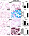
Fig 5. FOXM1 and AGR2 are highly expressed in human pulmonary invasive mucinous adenocarcinomas (PIMAs).
Representative images of lung tissue sections from patients with PIMAs (n = 12 patients, S2 Table). Images include adjacent normal lung tissue (left panels) and tumor lesions (right panels) in matched patients. Tissue sections were stained with antibodies against FOXM1, AGR2, NKX2.1 or stained for mucus using Alcian blue (left panels). Image J software was used to quantify intensity of staining (right graphs). A minimum of 5 random 20x field images per patient were quantified. Increased FOXM1 staining in PIMAs was associated with abundant Alcian blue staining, loss of NKX2.1 and increased expression of AGR2 in tumor cells.
Fig 6
Fig 6. FOXM1 induces AGR2 in human and mouse lung adenocarcinomas.
(A) In human PIMAs, immunofluorescence co-localization studies demonstrated that FOXM1-positive tumor cells were highly positive for AGR2. (B) In mouse urethane-induced lung cancer model, AGR2 was induced in epiFoxM1-ΔN tumors. Immunofluorescence staining for AGR2 in control and epiFoxM1-ΔN lung tumors (left panels, n = 6 control mice and n = 9 epiFoxM1-∆N mice, ten images per each mouse lung) and qRT-PCR of Agr2 mRNA using RNA from microdissected control (n = 5) and epiFoxM1-ΔN (n = 5) lung tumors (right panel). β-actin mRNA was used for normalization. A p-value <0.01 is marked with a double asterik (**). (C) In mouse KrasG12D –induced lung cancer models, AGR2 was induced in lung tumors of KrasG12D/ epiFoxM1-ΔN mice (n = 3) compared to control KrasG12D mice (n = 3). (D) FoxM1 is required for AGR2 expression in PIMAs. Human PIMA cell lines A549 and H2122 were stable transduced with control (scramble) or shRNAs against human FoxM1 (shFoxM1 #1 or shFoxM1 #2). Efficient inhibition of FoxM1 decreased Agr2 expression in both A549 (top) and H2122 (bottom) PIMA cells, shown with qRT-PCR. β-actin mRNA was used for normalization. A p-value <0.01 is marked with a double asterik (**). (E) Western blot shows the correlation of the loss of FOXM1 and AGR2 in the A549 (top) and H2122 (bottom) cell lines.
Fig 7
Fig 7. FOXM1 increases transcription of AGR2 and is required to maintain mucinous phenotype in PIMAs.
(A) Schematic of a potential FOXM1 binding site in the AGR2 promoter (left panel). ChIP shows binding of FOXM1 to the AGR2 promoter in A549 cells (right panel). A549 cells were fixed, lysed, sonicated, and used for immunoprecipitation with an antibody against FOXM1 or rabbit control IgG. PCR was performed encompassing a predicted FOXM1 binding site from -257bp to -247bp of the human AGR2 promoter. DNA region located at –6.0 kb of Agr2 promoter was used as a negative control. (B) FOXM1 transcriptionally activates AGR2 promoter. Sequence denotes the predicted binding motif and the sequencing results following mutagenesis (top). Dual luciferase assay in Hek293T cells transfected with the WT (AGR2-Luc) or mutated -2.0kb AGR2 (mut AGR2-Luc) promoter and an empty (CMV-Empty) or human FoxM1 expression (CMV-FoxM1) vectors is shown (bottom). A p-value <0.01 is marked with a double asterik (**). (C) Depletion of FOXM1 in human invasive mucinous A549 adenocarcinoma cells decreased lung tumor growth in the orthotopic xenograft model of lung cancer. A549 cells were inoculated into tracheas of immunocompromised mice. Representative images of H&E staining (5–8 images per mouse lung) and photographs of mouse lungs after eight weeks post tumor inoculation are shown (left panels). Tumor numbers and sizes in the two groups (n = 6 control (Scramble) and n = 6 shFoxM1 mice) are shown (right panel). (D-E) FoxM1 is required to maintain mucinous phenotype in human PIMAs shown in orthotopic xenograft model of lung cancer (n = 6 control mice and n = 6 shFoxM1 mice). (D) Inhibition of FOXM1 reduces mucus depositions shown with Alcian Blue, and (E) reduces AGR2 and MUC5AC expression in A549 orthotopic xenografts. AGR2 co-localizes with MUC5Ac in the orthotopic A549 xenografts. 5–8 images per mouse lung were used. (F) Frequency of observed mediastinal lymph node and liver metastases in control (Scramble) mice (n = 6) and shFoxM1 mice (n = 6). (G) Control (Scramble) orthotopic A549 xenograft tumors (green) invade airways (purple). No invasion of tumor cells into airways was found in FOXM1-deficient tumors.
Fig 8
Fig 8. FOXM1 stimulates progression of lung adenomas into mucinous adenocarcinomas.
Schematic drawing shows that FOXF1 induces expression of cell-cycle regulatory and mucinous genes, including Agr2, causing increased tumor cell proliferation and mucinous phenotype. FOXM1 directly activates transcription of Agr2. Both FOXM1 and AGR2 are critical for PIMA growth, invasion and progression of lung adenomas into aggressive mucinous adenocarcinomas.

References

    1. Travis WD, Brambilla E, Noguchi M, Nicholson AG, Geisinger K, Yatabe Y, et al. International Association for the Study of Lung Cancer/American Thoracic Society/European Respiratory Society: international multidisciplinary classification of lung adenocarcinoma: executive summary. Proc Am Thorac Soc. 2011;8(5):381–5. doi: 10.1513/pats.201107-042ST . - DOI - PubMed
    1. Jagirdar J. Application of immunohistochemistry to the diagnosis of primary and metastatic carcinoma to the lung. Arch Pathol Lab Med. 2008;132(3):384–96. doi: 10.1043/1543-2165(2008)132[384:AOITTD]2.0.CO;2 . - DOI - PubMed
    1. Maeda Y, Tsuchiya T, Hao H, Tompkins DH, Xu Y, Mucenski ML, et al. Kras(G12D) and Nkx2-1 haploinsufficiency induce mucinous adenocarcinoma of the lung. J Clin Invest. 2012;122(12):4388–400. doi: 10.1172/JCI64048 ; PubMed Central PMCID: PMC3533546. - DOI - PMC - PubMed
    1. Winslow MM, Dayton TL, Verhaak RG, Kim-Kiselak C, Snyder EL, Feldser DM, et al. Suppression of lung adenocarcinoma progression by Nkx2-1. Nature. 2011;473(7345):101–4. doi: 10.1038/nature09881 ; PubMed Central PMCID: PMC3088778. - DOI - PMC - PubMed
    1. Hwang DH, Sholl LM, Rojas-Rudilla V, Hall DL, Shivdasani P, Garcia EP, et al. KRAS and NKX2-1 Mutations in Invasive Mucinous Adenocarcinoma of the Lung. J Thorac Oncol. 2016;11(4):496–503. doi: 10.1016/j.jtho.2016.01.010 . - DOI - PubMed

Publication types

MeSH terms